Vascularization Of The Central Nervous System: Characteristics And Structure
Our brain needs to receive a constant and solid blood supply loaded with nutrients and oxygen, since it does not have the capacity to store the energy from the food we eat.
This is possible thanks to correct vascularization of the central nervous system (CNS) which is made up of a complex interconnection of arteries and blood vessels that are distributed throughout the spinal cord and brain.
The vascularization of the CNS allows, in addition to providing nutrients and oxygen to the brain, it and each of its parts can develop their functions.
Before detailing what the functions of the vascularization of the central nervous system consist of, we will comment, as a summary, on the types of arteries that make up the central nervous system.
The vascularization of the central nervous system (CNS) is possible thanks to the arteries that reach different areas that make up its structure.
To receive this blood supply, so necessary in the brain, there are two arterial groups that are responsible for it, coming from the heart and through the aorta artery, thus allowing the maintenance of the metabolic activity of the body.
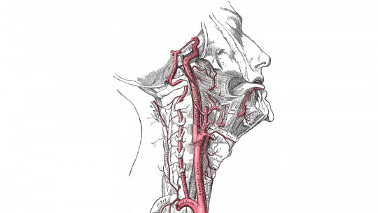
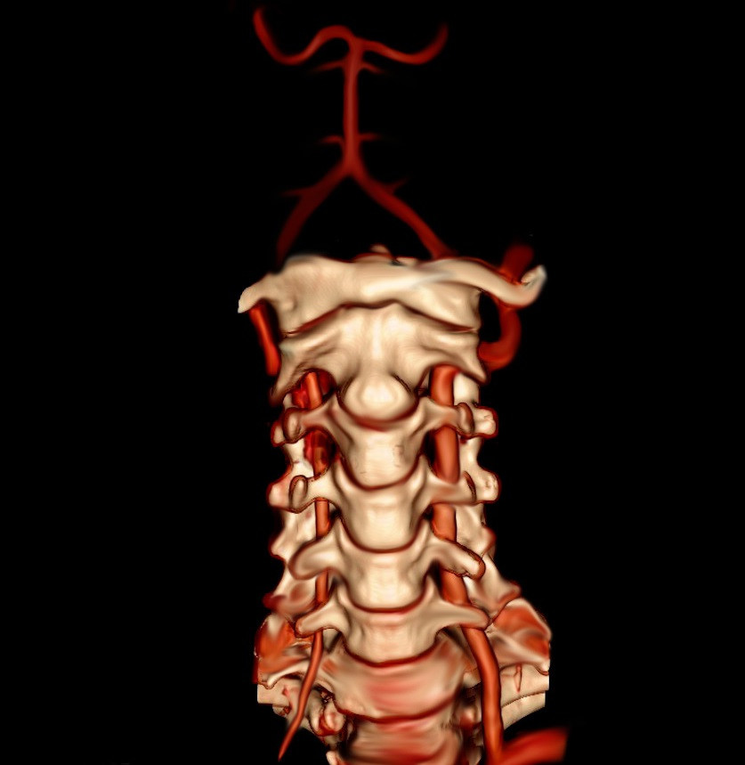
On one side are the vertebral arteries, which are responsible for supplying the caudal or posterior area of the brain; joining so that they make up the basilar artery which, in turn, forms the posterior cerebral artery. These are responsible for supplying blood to the brain stem and cerebellum.
On the other hand, there are the internal carotid arteries, which have the task of supplying the rostral or frontal area of the brain, forming the anterior and middle cerebral arteries. These arteries are divided into smaller branches that spread through the subarachnoid space or leptomeningeal space and enter the brain tissue in order to ensure that it is provided with the necessary nutrients for its proper functioning.
The aforementioned arteries can also be of two types. One type is made up of conduction channels, which are directed towards the lateral surfaces of the brain, and the other type is made up of perforating cells which come from the conduction arteries in order to supply more specific areas.
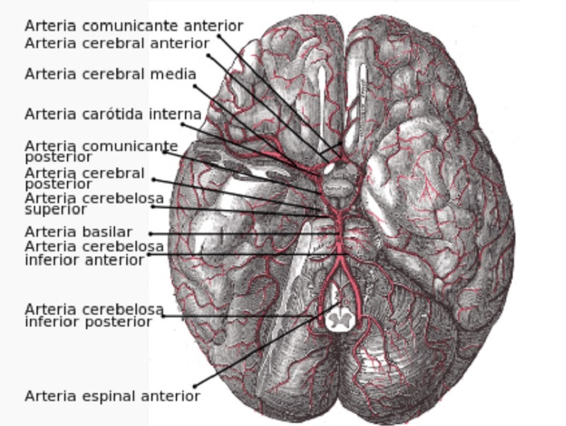
There is an area where the basilar artery and the carotid arteries connect, a structure called the circle of Willis, which is the area found in the lower part of the brain and in which the internal carotid arteries branch into smaller arteries. size, the latter being the ones that They are responsible for supplying oxygen-laden blood to 80% of the brain
Vascularization of the spinal cord
The area of the central nervous system, called the spinal cord, is divided into the following segments, like the vertebral column: cervical, thoracic, lumbar, sacral and coccygeal. Each and every one of these segments is responsible for providing eight pairs of spinal nerves that leave from the spinal canal.
For the correct functioning of the spinal cord and all its segments, correct vascularization through the arteries and venous ducts that cross it is essential, as will be explained in more detail below.
1. The irrigation of the arteries of the spinal cord
The spinal cord is the area of the central nervous system that is responsible for transmitting messages in and out from the brain to the rest of the body. Now, for correct operation, It is crossed by three arterial vessels longitudinally these being the anterior spinal artery and the two posterior spinal arteries.
This anterior spinal artery originates from the two vertebral arteries that are located at the level of the medulla oblongata, also known as the medulla oblongata, and goes down through the anterior or frontal surface of the spinal cord.
On the other hand, the posterior spinal arteries, which emerge from the vertebral arteries or the posterior inferior cerebellar arteries and depart towards the caudal or posterior surface of the spinal cord.
The arteries of the spinal cord, previously mentioned, need to be reinforced by the radicular arteries, such as the ascending cervical, intercostal and lumbar arteries, in order to be able to supply blood through the spinal cord, through the part located below of the cervical segments.
A disorder produced in the irrigation of the spinal cord of the CNS, such as an occlusion in the anterior spinal artery, causes what is known as “acute thoracic spinal cord syndrome” which entails paraplegia and incontinence, also losing sensitivity to temperature and blood pressure. pain.
2. Drainage of the venous ducts of the spinal cord
The drainage action of the venous conduits of the spinal cord occurs through a pattern similar to that of the arterial irrigation of this area. For it, There are six interconnected venous tubes that are expanded longitudinally by the spinal cord
These conduits constitute the anterior and posterior spinal veins; both extended to the middle area. On the other hand, there are the anterolateral veins and the posterolateral veins that are close to the insertion of the anterior and posterior venous roots.
The set of these blood vessels are responsible for draining, through the anterior and posterior radicular veins, to the epidural venous plexus also known as the internal vertebral venous plexus, which is located between the vertebral peristyle and the dura mater, which is the outer layer that is responsible for covering and protecting both the brain and the spinal cord.
In addition, the internal venous plexus is connected to the external vertebral venous plexus, so it is interconnected with the ascending veins of the lumbar area and with the azygos and hemiazygos veins, which fulfill a special function by providing an alternative route for blood circulation to the right atrium of the heart, if a situation arose in which the other chambers were blocked.
The part of the central nervous system known as the brain is made up of three main areas: the cerebrum, the cerebellum, and the brainstem. All these areas are at full capacity thanks to correct vascularization.
1. The irrigation of the arteries of the brain
The part of the central nervous system known as the brain is supplied by two pairs of blood vessels, better known as internal carotid arteries and vertebral arteries.
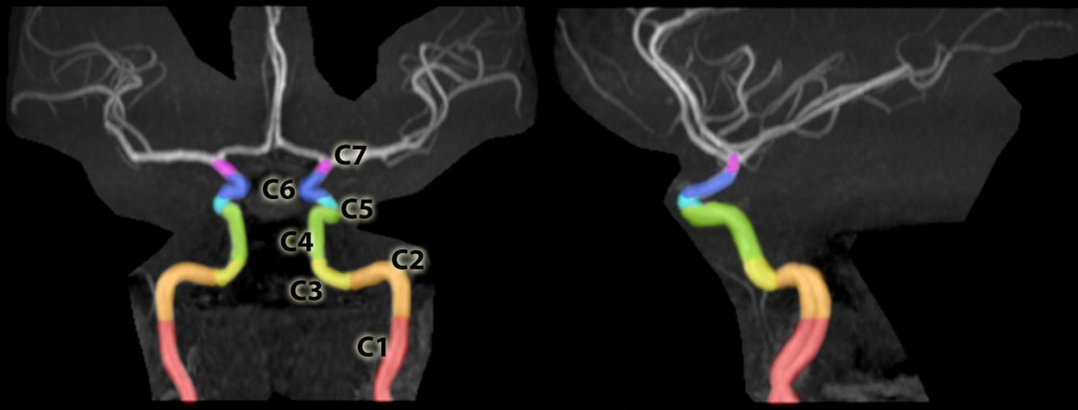
internal carotid artery
The internal carotid artery It is divided between two arteries known as the anterior and middle cerebral arteries
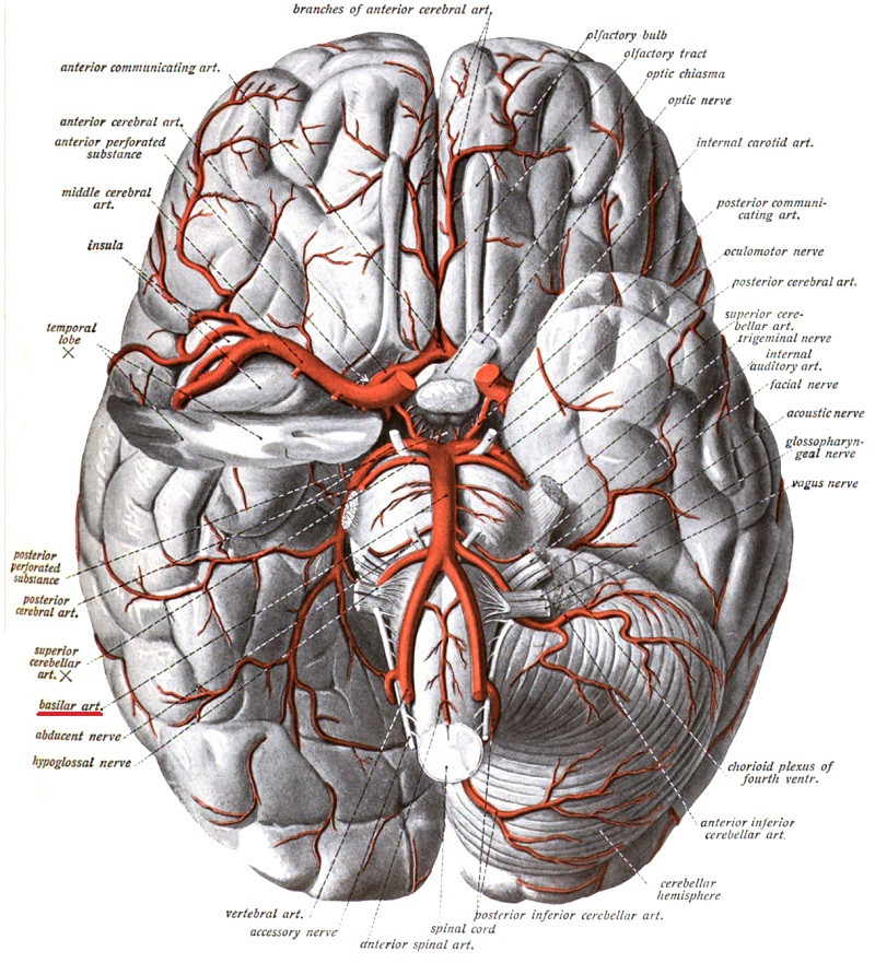
The anterior cerebral artery passes over the optic nerve and subsequently crosses the longitudinal fissure found between the two cerebral hemispheres, following the curvature of the corpus callosum, until it supplies the medial area of the frontal and parietal lobes. It also joins the blood vessel on the opposite side through the anterior communicating artery. This is why the anterior cerebral artery is responsible for supplying the brain areas of the motor and sensory cortex of the lower limb (the legs).
The middle cerebral artery, being the largest of the three arteries of the brain, has a larger cortical territory than the others. Starting from the place where it originates, it continues until it penetrates the lateral sulcus of the brain, where it is divided so that its branches are responsible for irritating the lateral area of the temporal, parietal and frontal lobes.
This entire surface covers the primary motor and sensory cortices of the entire body, with the exception of the lower limb. In addition, it is responsible for irrigating the auditory cortex and the insula, located deep in the lateral sulcus of the brain.
Vertebral artery
The vertebral artery arises from the subclavian artery rising towards the transverse foramina, located in the cervical vertebrae, until entering the cavity of the skull, passing through the foramen or foramen magnum.
In this journey, The vertebral artery branches into arteries called anterior and posterior spinal arteries which are responsible for supplying the spinal cord and medulla oblongata.
Among all these branches there is one branch that stands out above the rest as it is larger in size; It is known as the posterior inferior cerebellar artery whose function is to irrigate the lower part of the cerebellum.
When crossing the rostral or frontal area, the two vertebral arteries join in the area of the medulla oblongata, forming the basilar artery
This basilar artery is in turn branched so that it supplies multiple areas, including the lower and anterior parts of the brain, through the anterior cerebellar artery; also the inner ear, through the labyrinthine artery.
The basilar artery is also subdivided into the superior cerebellar arteries and the posterior cerebral arteries The superior cerebellar is responsible for supplying the upper layer of the cerebellum, while the posterior cerebellar has the task of supplying the inferomedial aspect of the temporal lobe, as well as the visual cortex of the occipital lobe.
In composition, the irrigation of the brain by the vertebral and basilar arteries has been called the “vertebrobasilar system.” All of this includes a vascular network located at the base of the brain, the aforementioned circle of Willis, also called the arterial circuit of the brain.
2. Drainage of the venous ducts of the brain
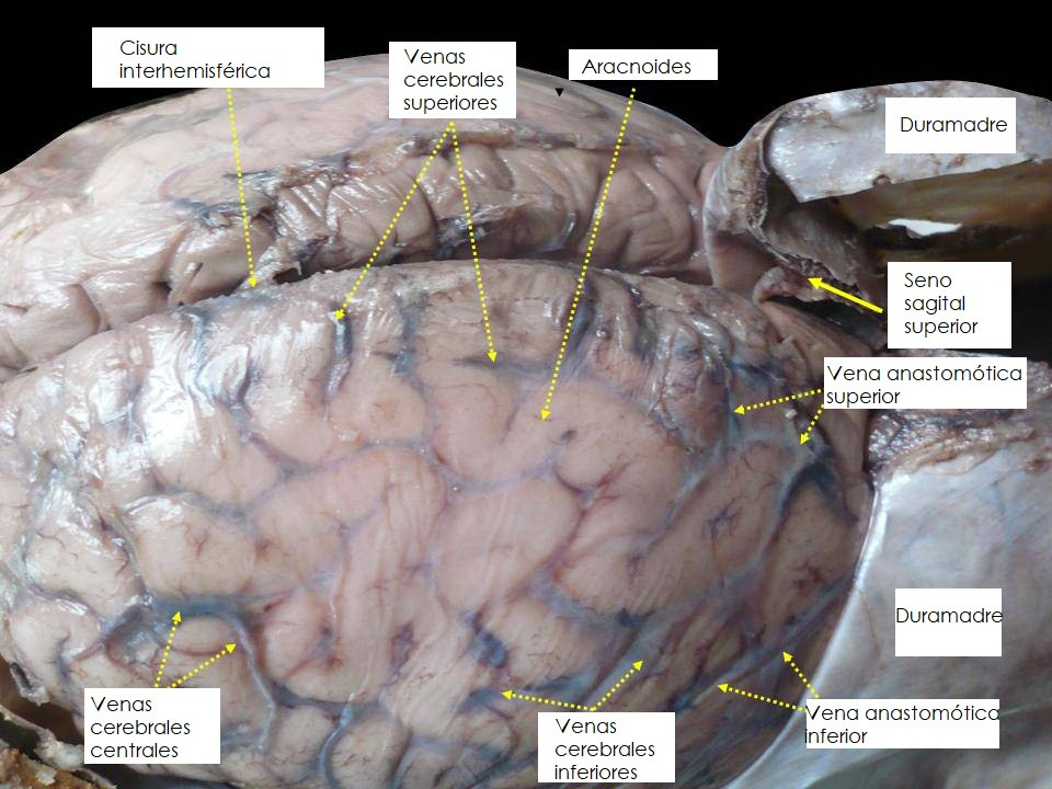
For the drainage of this part of the central nervous system there are three vessels that allow it: the venous sinuses, the superficial veins and the deep veins.
The deep and superficial cerebral veins are responsible for draining the venous sinuses located in the layer known as the dura mater, and are pathways that are formed between the two sheets of the dura mater and which in turn are subdivided between:
Superior sagittal sinus: responsible for receiving blood from the superior cerebral veins.
Inferior sagittal sinus: through which the veins located on the medial side of the hemispheres are drained.
Straight sinus: in which area the deeper structures of the forebrain are drained, in addition to the inferior sagittal sinus.
The deep cerebral veins, in turn, They fulfill the function of draining the structures located in the internal part of the forebrain It is worth highlighting the choroidal and thalamostriate veins, which are responsible for draining the thalamus, basal ganglia, hippocampus, choroid plexus and internal capsule.
These veins They join together to form the internal cerebral vein and, in addition, the two internal cerebral veins form the great cerebral vein or Galen’s vein located in the lower part of the corpus callosum, continuing through the straight sinus, located in the cerebellum and responsible for draining the internal jugular vein that receives blood from the face, neck and brain.
The superficial veins are located in the subarachnoid space and its function is to drain the lateral surface of both cerebral hemispheres, until reaching the superior sagittal sinus.
Damage to the vascularization of the central nervous system
A stroke occurs when the vascularization of the brain is interrupted, being what is equivalent in the brain to a myocardial infarction in the heart. This causes damage that may be irreversible in the person who suffers it.
As mentioned above, the brain needs to receive nutrients and oxygen through the circuit that makes up the vascularization of the central nervous system; Therefore, if this vascularization is interrupted, the brain cells begin to fail, even dying and potentially causing what is known as a stroke or cerebral infarction.
This occurs most of the time due to blockage of one of the blood vessels and, therefore, when there is a lack of oxygen, this hinders the correct physical and mental functioning of the person, which is why they can suffer serious damage. Likewise, with the help of professionals you can gradually recover the affected functions and even relearn skills.
The best-known habits to prevent a stroke are controlling blood pressure and cholesterol levels, as well as avoiding smoking.

: Structure and Functions of This Layer of the Meninges")
: Parts, Functions and Diseases")


: Parts and Functions")