

See more of our articles in your search results.
A car horn blares behind you while you stand at the crosswalk, and before you have thought a single word your heart has jumped, your head has turned and every sense has snapped to attention. Three hours later you are in a meeting, a perfectly comfortable chair under you, trying to stay awake while a colleague reads slides aloud. Same brain. Same person. One moment it is blazing; the next it is a dimmer switch somebody has left at a quarter. Something in your head decides, all day long, how awake you are and what deserves your attention, and you have almost no say in the matter.
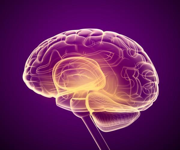
That something is largely the ascending reticular activating system, or ARAS, a network of nerve cell groups in the upper brainstem that sends signals up to the thalamus, hypothalamus, basal forebrain and cortex. It does not think, remember or decide. What it does is more basic: it keeps the thinking parts of your brain switched on. Without it, the rest of the brain can’t sustain wakefulness at all.
It is also one of the most misunderstood structures in popular science, and the confusion cuts both ways: some people ignore it, and others credit it with powers it doesn’t have.
This guide explains what the ascending reticular activating system is, its parts and functions, and how to activate it, in plain language and with the evidence behind each claim.
You’ll also find where social-media wisdom runs ahead of the science, what happens when the system is damaged, and when persistent drowsiness or overdrive deserves a doctor’s attention.
How many of these have you been "meaning to read" for months?
- 1
 TarcherThe Artist's Way30th Anniversary EditionListen Free On Audible
- 2
 Undercover Publishing HouseStop Letting Everything Affect YouHow to break free from overthinking, emotional chaos, and self-sabotage.Listen Free On Audible
- 3
 MajostaDon't Believe Everything You ThinkWhy Your Thinking Is The Beginning & End Of Suffering (Beyond Suffering)Listen Free On Audible
What is the ascending reticular activating system?
The ascending reticular activating system (ARAS) is a set of brainstem nuclei and their connections that regulate arousal: your level of wakefulness, alertness and readiness to respond. “Ascending” refers to its upward projections, “reticular” to the net-like core of the brainstem where it was first located, and “activating” to its role in switching the cortex into a wakeful state.
The idea comes from a classic 1949 experiment. Neuroscientists Giuseppe Moruzzi and Horace W. Magoun, working with cats, found that electrical stimulation of the brainstem reticular formation changed the brain’s electrical rhythm on the EEG. The slow, synchronized waves of a drowsy brain gave way to low-voltage, fast activity, the signature of an alert one. This effect was not carried by the known sensory pathways that run through the brainstem, which suggested a separate system dedicated to activation. Their paper in Electroencephalography and Clinical Neurophysiology became a landmark in neuroscience.
Before that, many scientists thought of sleep as the cortex simply running out of input. Moruzzi and Magoun’s work flipped the picture: wakefulness is actively generated by the brainstem, and the cortex needs that upward push to stay awake. Later studies showed that damage to this region can cause coma, even when the cortex itself is intact.
Modern researchers often prefer a slightly different term. Some now speak of the ascending arousal network (AAN), partly because many of the nuclei that contribute to arousal sit outside the classic reticular core, and partly because “network” better conveys that many modular circuits work together. You’ll still see ARAS widely used in textbooks and clinical writing, and this article uses both.
A word about scope. The ARAS regulates arousal, which is not the same as awareness. You can be highly aroused and confused, or calm and acutely aware. Consciousness depends on both the brainstem’s arousal systems and the cortical networks that generate the content of experience. The ARAS is the power supply, not the screen.
Think of Priya, 24, a graduate student who feels awake the instant she steps into a cold, bright lab after a night of five hours’ sleep. Part of what she’s feeling is her arousal system responding to light, temperature and novelty. Her thoughts, meanwhile, are another matter entirely.
In short: the ARAS is the reason you can read this sentence with some alertness instead of snoozing through it.
Where is the ARAS located, and what are its parts?
It lives mostly in the pons and midbrain of the brainstem, with extensions into the hypothalamus, thalamus and basal forebrain. It isn’t a single structure but a collection of cell groups, each releasing its own chemical messenger, which together project widely across the brain.
A 2012 study by neurologist Brian L. Edlow and colleagues used a technique called high angular resolution diffusion imaging to map the structural connectivity of the human ARAS in three adult brains, two examined after death. They identified the key source nuclei in the brainstem, including the cuneiform and subcuneiform nuclei, the pontis oralis, the median and dorsal raphe, the locus coeruleus, the pedunculopontine nucleus, the parabrachial complex and the ventral tegmental area. Their tractography found that these brainstem nuclei connect mainly with the intralaminar, paraventricular and reticular nuclei of the thalamus, and with the hypothalamus. The study also revealed pathways to the basal forebrain, and some that appeared specific to humans. The sample was very small, so the findings are best seen as a detailed first draft of the human map.
| Component | Main chemical messenger | Role in arousal |
|---|---|---|
| Locus coeruleus (pons) | Norepinephrine | Alertness, vigilance, response to stress and novelty |
| Dorsal and median raphe nuclei | Serotonin | Modulates wakefulness, mood and sleep cycles |
| Ventral tegmental area | Dopamine | Motivation, reward-driven arousal |
| Pedunculopontine and laterodorsal tegmental nuclei | Acetylcholine | Cortical activation in waking and REM sleep |
| Parabrachial complex and other reticular nuclei | Glutamate | Excitatory drive to the forebrain |
| Tuberomammillary nucleus (hypothalamus) | Histamine | Maintains wakefulness |
| Lateral hypothalamus | Orexin (hypocretin) | Stabilizes the waking state |
| Thalamus (intralaminar nuclei) | Relay | Passes arousal signals to the cortex |
Two broad routes carry the signal upward. One travels through the thalamus, which then drives widespread cortical activity. The other bypasses the thalamus through the hypothalamus and basal forebrain, spreading monoamines and acetylcholine directly across the cortex. Together they ensure that the entire cortex, not just one region, shifts into an alert mode.
The distribution of chemicals matters in practice. Many drugs that change alertness, such as stimulants, antidepressants, antihistamines and some sleep medicines, act on one or more of these messenger systems. That’s why a cold remedy can make you drowsy while a stimulant makes you wired: they are pushing different levers of the same network.
If you only remember one thing from the table, make it this: the ARAS is a team of chemically distinct players, not a single “alertness center.”
What are the main functions of the ARAS?
Its central job is to regulate arousal, which supports nearly everything else you do. Researchers usually group its functions into a few overlapping roles.
- Maintaining wakefulness: keeping the cortex in an active state so that you can perceive, think and act.
- Controlling sleep-wake transitions: switching arousal down for sleep and back up for waking, together with hypothalamic sleep circuits.
- Supporting attention and vigilance: adjusting how alert and responsive you are to what’s happening around you.
- Triggering the orienting response: turning attention toward sudden or novel stimuli, such as a loud noise or your name in a crowded room.
- Contributing to consciousness: providing the level of arousal that awareness depends on.
The orienting response shows the system at work. When something unexpected happens, sensory pathways send branches to the brainstem, which responds by raising cortical arousal within a fraction of a second. That is why you can doze on a train yet wake instantly when your station is announced, and why a parent can sleep through traffic but not through a baby’s whimper. The system is selective: it tunes its response to what matters to you.
It also adjusts arousal to context and demand. Facing a difficult problem or a threat, arousal rises; in a quiet, familiar setting, it falls. Hormones and body rhythms contribute, which is why alertness tends to dip in the early afternoon and peak in the late morning for many people.
What the ARAS doesn’t do is just as important. It doesn’t store memories, understand language or generate thoughts. Those depend on cortical and subcortical networks that the ARAS switches on and tunes. You could think of it as the conductor’s cue that starts the orchestra, not the music.
Picture Daniel, 47, a night-shift nurse. At 3 a.m. a monitor alarm sounds, and within a second he is fully alert and moving. At 3:20, with the ward quiet, he struggles to keep his eyes open. His arousal system is doing exactly what it should: raising and lowering the dial in response to demand. The trouble is only that the demand schedule runs against his body clock.
A fair caveat: much of what we know about these functions comes from animal studies and from lesion research in people. Human imaging of the ARAS is technically difficult, because the nuclei are small and deep, so descriptions of precise roles in healthy human brains remain partly inferred.

How does the brain switch between sleep and wakefulness?
Through a mutual-inhibition circuit often described as a flip-flop switch. In a 2005 review in Nature, neuroscientist Clifford B. Saper and colleagues described how the arousal systems, including the monoaminergic cell groups, and sleep-promoting neurons in the hypothalamus’s ventrolateral preoptic nucleus (VLPO) inhibit one another. When wake-promoting cells are active, they suppress the VLPO; when the VLPO fires, it suppresses the arousal cells. This arrangement produces sharp transitions between states and discourages the brain from lingering in between.
The model offers a tidy explanation for experiences like falling asleep suddenly, or waking up fully rather than gradually. It also explains why drowsiness can feel like a battle: as sleep pressure builds, the balance of the switch tips, and the arousal system loses its grip.
A third group of cells stabilizes the switch. Neurons in the lateral hypothalamus that contain orexin (also called hypocretin) excite the arousal nuclei and help keep you awake. Saper’s review noted that loss of these cells leads to narcolepsy, a condition marked by overwhelming daytime sleepiness and abrupt shifts between sleep and wake. In other words, the switch can flip too easily when its stabilizer is missing.
Two pressures push the switch. The homeostatic drive builds up the longer you stay awake, partly through chemicals such as adenosine accumulating in the brain. The circadian drive, coordinated by the brain’s master clock in the hypothalamus, promotes alertness at certain times of day and sleepiness at others. Their combination explains why you can feel wide awake at 10 p.m. after a short night, or wiped out at 3 p.m. despite a full night’s sleep.
The flip-flop model is a simplification, and it has been refined since 2005; the real circuitry includes many more cell groups, and REM sleep requires its own interacting systems. Still, it remains a useful framework in teaching and sleep medicine.
Imagine Ayesha, 31, a new mother sleeping in short bursts. Her partner says she wakes at the faintest sound and then sinks back in seconds. That is a switch tuned to a particular concern, pivoting quickly between states in response to her baby.
What might this mean for you? The longer you push through sleep pressure with stimulants, bright light and sheer will, the more your system must work against its own switch, and the less stable your alertness tends to be.
How does the ARAS shape attention, stress and focus?
By setting the gain on the brain’s responsiveness. Neuroscientist Gary Aston-Jones and Jonathan Cohen proposed in 2005 an influential account of the locus coeruleus-norepinephrine system, one of the major ARAS components. The locus coeruleus, a tiny cluster of cells in the pons, sends norepinephrine to most of the brain. In their adaptive gain theory, it operates in two modes that shape performance.
In phasic mode, the locus coeruleus fires in brief bursts in response to task-relevant events, which sharpens focus and supports efficient performance. In tonic mode, it maintains a higher baseline firing rate, which promotes distractibility and a readiness to disengage from the current task and explore alternatives. Optimal performance on a focused task is thought to occur at a moderate level of tonic activity combined with strong phasic responses. Too little arousal leaves you drowsy and slow; too much leaves you jittery and scattered.
That fits the old observation that performance follows an inverted U against arousal. A person with a mild level of stress may do well on a hard task, whereas high stress can disrupt it. Everyday experience supports it: a little pressure before a presentation sharpens you, while panic scatters your thoughts.
The theory is a model built largely from animal recordings and computational work, and some of its details remain debated. Human evidence is more indirect, drawing on pupil size, which tracks locus coeruleus activity, and on pharmacological studies. Treat it as a well-regarded framework rather than settled fact.
Applied to daily life, the model explains several familiar states. Exhaustion produces low arousal. Anxiety can produce a high-tonic state in which attention jumps between threats. Flow and deep concentration correspond to the sweet spot. Some researchers have linked altered arousal regulation to attention difficulties such as ADHD, but the relationship is complex and not a simple matter of too little or too much.
Take Miguel, 36, an exam invigilator who finds that his best focus comes after a brisk walk and a medium coffee, and that a third coffee leaves him restless and checking the clock. He has discovered the shape of the curve without needing the theory.
The practical lesson: more arousal is not always better. The goal is the right level for the task.

What happens when the ARAS is damaged?
Serious damage to the brainstem arousal network can cause coma, a state in which a person can’t be awakened and shows no signs of awareness. Because the ARAS provides the upward drive that keeps the cortex active, lesions in the upper pons and midbrain can switch off wakefulness even if the cortex is healthy.
The same ARAS circuits are central to the clinical study of disorders of consciousness: coma, the vegetative state or unresponsive wakefulness syndrome, and the minimally conscious state. Neurologist Nicholas D. Schiff proposed in 2010 the mesocircuit hypothesis, which suggests that after severe brain injury, widespread loss of excitatory input to the central thalamus and its connections reduces the activity of frontal and striatal circuits, and that recovery depends partly on restoring this balance. His framework has helped guide research into treatments, including medications and deep brain stimulation, though evidence for specific therapies remains limited and mixed.
Not all arousal problems reflect structural damage. Common medical conditions can alter arousal, such as obstructive sleep apnea, narcolepsy, medication effects, thyroid disease, severe depression and certain infections. Sedatives and general anesthetics act, in part, by dampening the arousal systems. In the other direction, stimulants and some withdrawal states can overdrive them.
Because the ARAS is so closely tied to consciousness, doctors pay close attention to patients’ level of arousal, using tools such as the Glasgow Coma Scale. A sudden change in alertness, confusion or unresponsiveness is a medical emergency.
This is the point to be clear about what the article can and can’t do. It describes how the system works in general. It can’t evaluate your symptoms. If you or someone near you has sudden drowsiness that can’t be roused, new severe confusion, repeated fainting or signs of stroke, call local emergency services without delay.
Consider Hannah, 58, whose husband found her unusually difficult to wake one morning, mumbling and unable to answer simple questions. An ambulance was called, and the diagnosis, made in hospital, was a stroke affecting the brainstem. The speed of the response mattered more than any explanation of the anatomy.
Recovery from disorders of consciousness varies greatly, depending on the cause, the extent of injury and time. Specialist neurological and rehabilitation teams manage these cases, and families deserve honest, careful communication about uncertainty.
Does the reticular activating system really filter the world for your goals?
Not in the way the internet claims. Myth: your reticular activating system is a goal-detector that, once you write down what you want, filters reality to show you opportunities. Fact: the ARAS regulates arousal, and the idea that it acts as a manifestation filter has no direct scientific support.
Where does the myth come from? A kernel of truth. Attention is selective, and what you care about does shape what you notice. Buy a red car and you start seeing red cars; your name cuts through a noisy party. That experience, sometimes called the Baader-Meinhof phenomenon or frequency illusion, reflects attention and memory, working in cortical networks, not a goal-sorting function of the brainstem.
The ARAS contributes in a more basic way. It sets the level of arousal that makes selective attention possible and helps trigger the orienting response to significant stimuli. But the decisions about what is significant, whether it’s your name, a danger or a job posting, rely on higher brain areas that learn what matters to you.
Self-help writers have stretched the idea. They say that visualizing a goal “programs” the RAS to find it. That is a leap from the evidence. Goal-setting itself has decent research support: specific, challenging goals tend to improve performance. But the benefit comes through attention, planning and motivation across the brain, not through a hidden brainstem filter.
Here’s why it matters beyond pedantry. Believing that thinking about success will rewire a brainstem system can lead people to substitute positive thinking for action. Research on mental contrasting and implementation intentions suggests that pairing a goal with a realistic plan works better than visualizing success alone.
Think of Carlos, 29, who spent months visualizing a promotion while avoiding the uncomfortable conversation with his manager. His “RAS” was not the problem. A concrete plan, with a date, would have helped more.
You can enjoy the metaphor without swallowing the biology. Noticing opportunities is a real skill, and it can be trained, just not by the part of the brain this myth names.

How can you activate your ascending reticular activating system?
You can’t switch it on directly, but you can influence the conditions that drive it: sleep, light, movement, chemistry and context. The list below is based on what is known about arousal regulation; evidence is stronger for some items than for others, and I’ll say which.
- Protect sleep regularity. The strongest lever is a consistent sleep-wake schedule that gives your arousal system a clean baseline. Chronic sleep loss makes the system less stable, regardless of tricks. Evidence: strong.
- Get bright light early. Morning light helps anchor the circadian clock, which shapes alertness for the rest of the day. Ten to thirty minutes outdoors is a practical target. Evidence: good for circadian effects.
- Move your body. A brisk walk or short bout of exercise tends to raise alertness within minutes and improve mood and attention. Evidence: good for short-term effects.
- Use caffeine on purpose. Caffeine works mainly by blocking adenosine receptors, as Bertil B. Fredholm and colleagues described in a comprehensive 1999 review, which reduces the sleep-promoting signal and increases alertness. Time it for when you need it and avoid late-afternoon doses that can cut into sleep. Evidence: strong.
- Change the stimulus. Novelty, music, a change of scenery, a conversation or a standing posture can all raise arousal modestly. Brief cold water on the face or a short cool shower may also feel alerting, though research on lasting effects is limited. Evidence: modest.
- Take smart breaks. A short nap of about 10 to 20 minutes can restore alertness for many people, if taken early enough in the afternoon not to disturb nighttime sleep. Evidence: good.
- Cut the late-night glow. Evening light, especially from screens, can delay sleep and blunt next-day alertness. Evidence: moderate.
The last point has a specific study behind it. In a 2015 experiment, Anne-Marie Chang and colleagues compared reading on a light-emitting e-reader with reading a printed book before bed. After several nights, people using the e-reader took longer to fall asleep, had reduced evening melatonin, showed delayed circadian timing and felt less alert the next morning. The sample was small and the setting a sleep lab, but the finding fits the broader picture that evening light can interfere with the body clock.
Some popular “activation” tips, such as cold plunges, special breathing routines, binaural beats or supplements, have thin or mixed evidence. They aren’t necessarily harmful, but treat bold claims with skepticism, and remember that stimulating arousal beyond a moderate level can backfire. If you’re tired and wired, more activation isn’t the answer.
It also helps to match the method to the problem. If you’re sleepy because you slept badly, the fix is sleep. If you’re sluggish after a long meeting, movement and light may do it. If you can’t concentrate because you’re anxious, you may need to lower arousal, not raise it.
Take Joon, 33, who felt foggy by 2 p.m. every day. He moved his coffee to before noon, took a ten-minute walk after lunch and stopped scrolling in bed. Three weeks later, the slump had shrunk. The change was not dramatic; it was dependable.
What lowers arousal, and what overstimulates the system?
Sleep loss, sedating medications and monotony push arousal down, while stress, stimulants and constant interruption push it up. Many people are caught at both ends at once: exhausted by day, wired at night.
On the low side, the biggest culprit is insufficient or fragmented sleep. Sleep apnea, shift work, alcohol, some allergy and anxiety medications, depression and thyroid disease can all leave people drowsy and slow. A long, heavy meal or a warm, quiet room can also lower arousal. Boredom does so too; an unstimulating task may produce drowsiness in anyone.
On the high side, chronic stress activates the same systems you’d use to face a threat. The locus coeruleus fires more, stress hormones rise, and sleep gets lighter. Stimulants, high caffeine intake, nicotine, and certain medications raise arousal. Even constant notifications can keep the system on guard. Over time, a state of hyperarousal, with racing thoughts, difficulty falling asleep and an exaggerated startle response, is associated with insomnia, anxiety disorders and post-traumatic stress, though the link varies from person to person.
That brings up an overlooked point: calming the system matters as much as activating it. Techniques with decent evidence for lowering arousal include regular exercise earlier in the day, a wind-down routine, slow breathing, mindfulness practices, limiting late caffeine and alcohol, keeping the bedroom cool and dark, and cognitive behavioral therapy for insomnia, which is considered a first-line treatment.
Keep in mind that arousal isn’t purely good or bad. Both underarousal and overarousal can interfere with performance, and the right level depends on the task. Fine motor work and complex problem solving usually go best at moderate levels; simple, high-energy tasks tolerate more.
Picture Fatima, 44, a project manager who spends her day on caffeine and her evenings on her laptop. At 11 p.m. she is exhausted but can’t fall asleep. Her arousal system has been pushed up all day and has no off ramp. Her solution wasn’t more willpower: she moved her last coffee to noon, stopped work emails after eight and added a short walk after dinner.
If tiredness or wiredness persists for weeks despite good habits, a clinician can check for treatable causes.

When should you see a doctor about drowsiness or overdrive?
When it persists, interferes with daily life or comes with other worrying symptoms. Everyone has off days, but persistent changes in alertness can signal sleep disorders, medical conditions or mental health problems that respond well to treatment.
Signs that merit prompt evaluation include:
- Excessive daytime sleepiness for more than a few weeks despite adequate sleep, or falling asleep at inappropriate times such as during conversation or while driving.
- Loud snoring with pauses in breathing, morning headaches or waking unrefreshed, which may indicate sleep apnea.
- Sudden muscle weakness with strong emotions, vivid hallucinations at sleep onset or sleep paralysis, which can accompany narcolepsy.
- Persistent hyperarousal, such as chronic insomnia, constant tension or an exaggerated startle response after a frightening event.
A primary care doctor is a sensible starting point and can arrange blood tests, review medications and refer you to a sleep specialist, neurologist or mental health professional. A sleep study may be recommended for suspected sleep apnea or narcolepsy. For insomnia and hyperarousal tied to anxiety or trauma, a therapist trained in cognitive behavioral therapy or trauma-focused treatment can help.
Seek emergency help right away for sudden inability to wake someone, new severe confusion, a seizure, signs of stroke such as face drooping, arm weakness or slurred speech, or head injury followed by drowsiness. Contact local emergency services in those situations rather than waiting to see if things improve.
Be careful with self-diagnosis. The internet tends to turn ordinary tiredness into brainstem disease. A hard week, a poor night or too much coffee more commonly explains fluctuations in alertness than any disorder of the arousal system.
Imagine Samuel, 52, who laughed off his tiredness for two years. His wife kept noticing his loud snoring and pauses in breath. A sleep study showed severe sleep apnea, and with a breathing device his afternoons returned. The treatment was not exotic; the delay was.
The aim of understanding the ARAS isn’t to optimize every minute of alertness. It is to recognize when your wake system is being asked to do too much, or not enough.
FAQs about the Ascending Reticular Activating System
What is the ascending reticular activating system in simple terms?
It is a network of nerve cell groups in the upper brainstem that keeps the rest of your brain awake and alert. It sends signals upward to the thalamus, hypothalamus, basal forebrain and cortex, using chemical messengers such as norepinephrine, serotonin, dopamine, acetylcholine and histamine. It regulates arousal rather than thinking, memory or emotion. It was first described in 1949 by Giuseppe Moruzzi and Horace Magoun, who showed that stimulating the brainstem changed the brain’s electrical rhythm from a drowsy to an alert pattern. Damage to it can cause coma.
What are the parts of the ARAS?
The system includes several brainstem nuclei, including the locus coeruleus, the raphe nuclei, the pedunculopontine and laterodorsal tegmental nuclei, the parabrachial complex and the ventral tegmental area. It also involves the thalamus, particularly its intralaminar nuclei, the hypothalamus with histamine and orexin neurons, and the basal forebrain. Each group uses a different chemical messenger, and together they project widely across the cortex. A 2012 imaging study of three human brains mapped many of these connections, so the human picture is still being refined.
What does the ARAS do?
Its main role is to regulate arousal, which means your level of wakefulness and alertness. It helps keep the cortex active during waking, takes part in sleep-wake transitions, supports attention and vigilance, and triggers the orienting response when something sudden or novel happens. It also contributes to consciousness by supplying the level of arousal that awareness depends on. It does not store memories, understand language or generate thoughts, which rely on other networks that the ARAS switches on. Think of it as the power supply to the brain’s thinking machinery.
Can you really activate the reticular activating system?
Not directly, but you can influence the conditions that regulate it. Regular sleep, early daylight, movement, well-timed caffeine, short naps, novelty and avoiding late-night screens all affect alertness. Evidence is strongest for sleep, caffeine and light, and weaker for popular tricks such as cold exposure or special breathing. It also helps to match the method to the cause, since sleepiness from poor sleep needs sleep, not stimulation. If you are anxious and wired, lowering arousal may help more than raising it.
Does the RAS filter reality to help you achieve your goals?
There is no direct scientific support for this popular claim. Attention is selective, and what you care about does influence what you notice, as when your name cuts through a noisy room, but that selectivity depends on cortical networks that learn what matters to you. The ARAS sets the level of arousal that makes attention possible. Goal-setting works, but through planning and motivation, not a hidden brainstem filter. Pairing goals with specific plans is generally more effective than visualization alone.
What happens if the ARAS is damaged?
Severe damage to the brainstem arousal network, from stroke, bleeding, trauma or tumor, can cause coma, a state in which a person cannot be awakened. Milder or partial damage may lead to reduced alertness, excessive sleepiness or disturbed sleep-wake cycles. Researchers such as Nicholas Schiff have studied how circuits involving the thalamus contribute to recovery of consciousness after brain injury, and treatment research is ongoing. Sudden drowsiness that cannot be roused, new confusion or signs of stroke are emergencies that need urgent medical care.
Is the ARAS the same as the reticular formation?
They overlap but are not identical. The reticular formation is a diffuse network of neurons in the core of the brainstem, involved in arousal as well as breathing, heart rate, muscle tone and pain modulation. The ARAS refers to the ascending arousal components. Researchers increasingly use the term ascending arousal network because many of the key nuclei, such as the locus coeruleus and parabrachial complex, lie outside the classic reticular core and because many circuits work together. You may see the terms used loosely in textbooks and articles.
Why do I feel sleepy in the afternoon even after a good night’s sleep?
Two forces explain much of it. Your body clock produces a natural dip in alertness in the early afternoon, and sleep pressure, driven partly by adenosine, builds the longer you are awake. Large lunches, warm rooms and boring tasks can add to it. Strategies such as light exposure, a short walk, a brief nap of 10 to 20 minutes or caffeine earlier in the day can help. If you are sleepy every day despite enough sleep, snore heavily or doze at inappropriate times, a doctor can check for conditions such as sleep apnea.
Bibliography
- Aston-Jones, G., & Cohen, J. D. (2005). An integrative theory of locus coeruleus-norepinephrine function: Adaptive gain and optimal performance. Annual Review of Neuroscience, 28, 403-450.
- Chang, A.-M., Aeschbach, D., Duffy, J. F., & Czeisler, C. A. (2015). Evening use of light-emitting eReaders negatively affects sleep, circadian timing, and next-morning alertness. Proceedings of the National Academy of Sciences, 112(4), 1232-1237.
- Edlow, B. L., Takahashi, E., Wu, O., Benner, T., Dai, G., Bu, L., Grant, P. E., Greer, D. M., Greenberg, S. M., Kinney, H. C., & Folkerth, R. D. (2012). Neuroanatomic connectivity of the human ascending arousal system critical to consciousness and its disorders. Journal of Neuropathology & Experimental Neurology, 71(6), 531-546.
- Fredholm, B. B., Bättig, K., Holmén, J., Nehlig, A., & Zvartau, E. E. (1999). Actions of caffeine in the brain with special reference to factors that contribute to its widespread use. Pharmacological Reviews, 51(1), 83-133.
- Moruzzi, G., & Magoun, H. W. (1949). Brain stem reticular formation and activation of the EEG. Electroencephalography and Clinical Neurophysiology, 1(1-4), 455-473.
- Saper, C. B., Scammell, T. E., & Lu, J. (2005). Hypothalamic regulation of sleep and circadian rhythms. Nature, 437(7063), 1257-1263.
- Schiff, N. D. (2010). Recovery of consciousness after brain injury: A mesocircuit hypothesis. Trends in Neurosciences, 33(1), 1-9.
Use this citation format to reference the article clearly and help readers find the original source.
PsychologyFor. (2026). Ascending Activating Reticular System: Characteristics, Parts and Functions. PsychologyFor. https://psychologyfor.com/ascending-activating-reticular-system-characteristics-parts-and-functions/