The inferior temporal gyrus is a brain structure located in the lower part of the temporal lobe and an area that we know is responsible for visual perception and discrimination, as well as other functions that have been investigated later, such as arithmetic and mathematical processing. the numbers.
In this article we explain what the inferior temporal gyrus is what its structure and location, what functions it performs in the brain and what are the main disorders associated with damage to this brain structure.
Inferior temporal gyrus: definition, structure and location
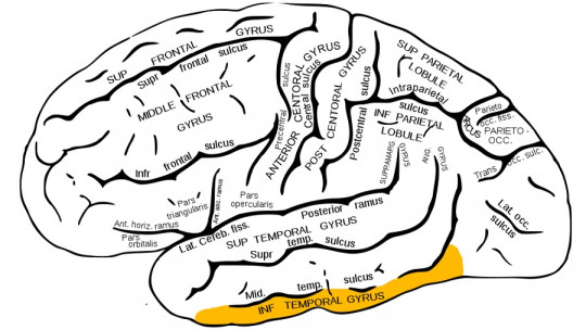
The inferior temporal gyrus is a gyrus of the brain located, as its name suggests, in the lower part of the temporal lobe This lobe consists of three gyri on its lateral surface: the superior, the medial and the inferior, which we will talk about throughout the article.
The gyri or gyri are the folds that give that wrinkled appearance to the cerebral cortex, the most developed area of the brain and the one responsible for higher cognitive functions such as thinking, language, planning or decision making.
The inferior temporal gyrus is, as we mentioned, one of the three gyri of the temporal lobe, one of the 6 main lobes that make up each cerebral hemisphere and whose main functions involve the management of auditory language, participation in the understanding systems of the speech, visual recognition of objects or identification of faces, among others.
The inferior temporal gyrus is located at the lower lateral edge of each cerebral hemisphere. below the medial temporal gyrus and behind the inferior occipital gyrus It extends around the inferolateral border to the inner surface of the temporal lobe, where it is limited by the inferior sulcus.
It should also be noted that the main source of blood supply to this brain area comes from the four temporal branches of the middle cerebral artery that emerge from the lateral sulcus or Sylvian fissure, a slit that crosses the entire brain transversely from its base and through. both sides.
Features
The inferior temporal gyrus It is a cerebral gyrus that is involved in visual object recognition and visual image processing due to its connection with areas of the inferior occipital gyrus that form the occipital lobe, the main brain structure related to visual perception and the interpretation and recognition of images, as well as spatial recognition or the discrimination of movements and colors.
The inferior temporal gyrus also appears to be specialized in interpreting and processing numbers In a study carried out in Palo Alto (United States), it was found that the inferior temporal area was activated in a differentiated and significant way when participants were presented with different numbers and their digits (e.g. “45” or “9”). ), which did not happen if the same was done with letters (“forty-five” or “nine”) or with homophonous words (e.g. “Hun” instead of “1”).
Although it was already known that this area of the brain was involved in the processing of visual information, with this and other research it was possible to reach the conclusion that this region was also involved in the arithmetic processing of numbers.
On the other hand, in another study carried out in Japan using functional magnetic resonance imaging, it was found that the left inferior temporal gyrus played a an important role in writing logograms (system of characters or signs that alone represent a meaning, used in languages such as Chinese) and, by extension, in other non-alphabetic languages.
Disorders related to damage to this structure
Lesions in a brain structure such as the inferior temporal gyrus can cause disorders such as visual agnosia, which involves an inability to identify, recognize and interpret visual stimuli. When unilateral damage occurs, the functional specialization of the cerebral hemispheres is revealed: only lesions on the right side affect higher visual functions. And why does this happen?
In most people, the left hemisphere is dominant when it comes to language, as well as the elaboration and interpretation of visual stimuli; while the right or non-dominant hemisphere would be specialized in non-verbal material and information, as occurs with the recognition of faces and emotional facial expressions. That is why only higher visual functions are affected when damage occurs to the right temporal area.
Another common disorder when a person suffers damage to the inferior temporal gyrus and adjacent structures is prosopagnosia or face blindness which causes an inability to recognize and discriminate faces, which can also lead to problems doing the same with objects or places.
Several studies have also concluded that damage to structures of the temporal lobe, specifically in the inferior and medial temporal area, can cause problems related to semantic memory, responsible for recognizing the meaning of objects, vocabulary or general knowledge. This is what is observed in semantic dementia, a neurodegenerative disease characterized by the gradual loss of this type of memory, both at a verbal and non-verbal level.
Finally, it should be noted that in some investigations it has been possible to conclude that lesions in the inferior temporal gyrus would result in an agraphia of logographic writing systems, such as kanji (logograms used in the Japanese language) or Chinese characters. Agraphias affect the person’s ability to write correctly and usually occur together with other language disorders such as aphasia and alexia.
Clark, DL, Boutros, NN, & Méndez, MF (2012). The brain and behavior: neuroanatomy for psychologists. Modern Manual. Nobre, A.C., Allison, T., & McCarthy, G. (1994). Word recognition in the human inferior temporal lobe. Nature, 372(6503), 260-263. Snell, R.S. (2007). Clinical neuroanatomy. Pan American Medical Ed.