Spinal Ganglia: Anatomy, Characteristics And Function
The spinal ganglia are a group of nodules located in the dorsal or posterior roots of the spinal nerves, where they are housed. the bodies of the neurons of the afferent or sensory pathway of the peripheral nervous system
In this article we will explain what the spinal ganglia are and their relationship with each of the parts of the peripheral nervous system.
The peripheral nervous system (PNS) includes the spinal nerves, cranial nerves, and their associated ganglia (groups of nerve cells outside the central nervous system (CNS)). Nerves contain nerve fibers that conduct information to (afferent) or from (efferent) the CNS
Generally, efferent fibers participate in motor functions, such as muscle contraction or gland secretion; and afferent fibers transmit sensory stimuli from the skin, mucous membranes and deep structures.
The main task of the PNS is to connect the various stimuli that our body receives (external, internal and proprioceptive or related to information about the position of one’s muscles) with the central nervous system; and the latter, in turn, connects with the organs and body systems that it must regulate and manage.
The PNS is made up of 12 pairs of cranial nerves, which exit the skull through several openings, and 32 pairs of spinal nerves, each of them identified by its relationship with the vertebra or spinal canal from which it emerges
spinal nerves
Spinal nerves extend from the spinal cord, passing through the vertebral muscles, to different areas of the body.
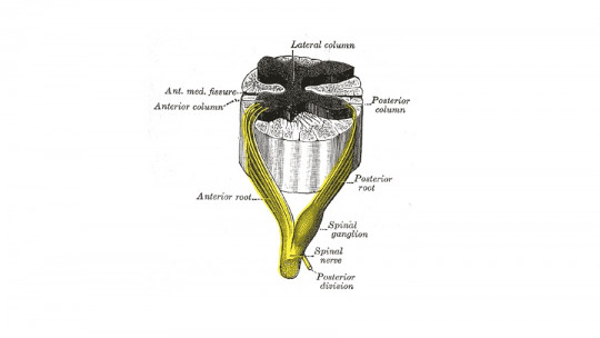
Each of the 31 pairs of spinal nerves has a ventral and a dorsal root ; Each root is made up of 1 to 8 tiny rootlets or bundles of nerve fibers. At the dorsal root of a typical spinal nerve, near the junction with the ventral root, is a dorsal or spinal root ganglion, a bulge containing nerve cell bodies.
The dorsal (or posterior) roots are primarily sensory. Each dorsal nerve root (except, in general, C1) contains afferent fibers (sensory or receptor) from the nerve cells of its ganglion. The dorsal roots contain fibers that come from cutaneous and deep structures.
Nerve fibers can be classified according to their anatomical and physiological basis in: efferent somatic fibers, which innervate skeletal muscles; and somatic afferent fibers, which transmit sensory information from the skin, joints, and muscles to the central nervous system.
The cell bodies of the afferent fibers are unipolar cells (characterized by having a single protruding extension of the soma) in the spinal ganglia, which are interposed in the course of the dorsal roots (dorsal root ganglia).
The peripheral branches of these ganglion cells are distributed throughout the somatic structures; and the central branches transmit sensory impulses through the dorsal roots to the dorsal cord of the gray matter and the ascending tracts of the spinal cord.
spinal ganglia
Nerve ganglia are groups of cells that make up small nodules located outside the central nervous system that function as relays or intermediate connections between different neurological structures of the body.
They can be divided into two types: the vegetative ganglia, made up of multipolar nerve cells located around the viscera on which they act, receive signals from the central nervous system and send them to the periphery (efferent function); and the spinal ganglia or dorsal root ganglia, made up of abundant distinctive neuronal connections, which are responsible for receiving signals from the periphery to send them to the brain (afferent function).
Spinal ganglia collect and modulate sensory information and constitute from a functional point of view the deposits of the neuronal somata of the primary afferent fibers of the entire sensory system, having specialized in higher animals as organs located outside the central nervous system.
The spinal ganglia group includes the spinal ganglia and the trigeminal (or Gasser), facial (or geniculate), glossopharyngeal (extracranial or Andersch and intracranial or Ehrenritter) and vagus (jugular and nodose) ganglia. .
The VIII pair or statoacoustic nerve also has two ganglia the vestibular or Scarpa and the cochlear, spiral or Corti, but their bipolar type neurons correspond to second-order neurons of a specialized sensory pathway whose functional meaning is not exactly similar to that of the general sensory or spinal ganglia.
Spinal ganglia injuries
Involvement of the spinal ganglia or dorsal roots can occur for various reasons among the most common we can find the following:
Herpes Zoster Infection
It is characterized by the appearance of localized, unilateral, imprecise pain, which precedes a vesicular eruption (appearance of vesicles or blisters on the skin) by 3 or 5 days. It can be accompanied by systemic symptoms such as fever, fatigue or myalgia
Vertebral tumors
They can produce, in addition to root injuries, other manifestations such as low back pain, pathological fractures, reduction in mobility or deviations of the spine. There are also primary tumors (neurofibroma) and metastatic tumors, such as lymphoma or meningeal sarcomatosis, in which several roots are usually affected.
Spinal ganglia and pain transmission
The sensation of pain appears when specific nerve fibers (called “A delta” and “C”) are activated. This activation can be triggered by poor functioning of the muscles and other soft tissues (which is what happens in “nonspecific syndromes”), or by various structural alterations that have been shown to be the cause of pain in some cases.
When stimulated, these nerve fibers They activate nerve cells in the spinal cord that transmit pain to the brain The nerve fibers “A delta” and “C” or sensitive to capsaicin, are thin and very numerous, and arise from the spinal ganglia, where their cell body is, bifurcating into two extremities.
When these nerve fibers are activated, they release substances (neurotransmitters or neuromodulators) that trigger inflammation of the innervated tissues. That inflammation triggered by the release of substances contained in the nerves (rather than by substances released by blood cells or tissues, as inflammation was classically understood) It is called “neurogenic inflammation.”
This type of inflammation can induce blood cells (such as macrophages, for example) to release substances that trigger classic inflammation (such as histamine), so that both types of inflammation would enhance each other. And in fact, the release of chemical mediators of inflammation could also increase or directly trigger pain.

: Parts and Functions")


