Brodmann’s 47 Areas, And The Brain Regions They Contain
Our brain is a large and complex set of organs, made up of different structures which fulfill different functions in the body. The most visible of all of them is the most external and developed in the human being, allowing its existence from the processing of information from the senses to the implementation of complex cognitive abilities: the cerebral cortex.
But the cerebral cortex does not have the same structure throughout its surface nor does it have the same functions at all its points. This has meant that for the study, imaginary divisions have been generated that limit different regions of the cortex. And of all the existing ones, The best known and most used is the Brodmann areas
The set of imaginary divisions into which the cerebral cortex can be divided and that allow the identification of specific regions of it are called Brodmann’s areas.
This division was proposed by the psychiatrist Korbinian Brodmann in 1909, the divisions not being random but based on the existence of differences regarding the composition, structure and arrangement of cells in different areas of the cortex.
The author’s objective was to generate a topographic classification from the study of anatomical characteristics, focusing on cytoarchitectonics and generating spatial divisions of the cortex to develop theory and be able to apply it in the field of pathology. The studies were carried out with a large number of animal species, but would only describe in detail the human brain and that of other apes
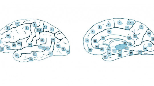
In the case of human beings, Brodmann divided them into a total of 47 zones or areas, although some of them can be subdivided to form a total of 52.
The Brodmann areas currently form a classification of brain areas best known and used worldwide having allowed mapping of the brain and being very useful both when investigating specific regions and when directing and carrying out different medical interventions.
The different areas of Brodmann
In the original classification, Brodmann divided the cerebral cortex into up to 47 different areas Specifically, we can find the following areas.
Brodmann Area 1
Brodmann area 1 can be found just after the central sulcus or Rolando fissure. It is part of the primary somatosensory area and works with somesthetic information coming from the body.
This area is also part of the primary somatosensory cortex, having the same functions as the previous one.
Brodmann Area 3
Along with the previous two, it is part of the primary somatosensory cortex. It also collects and processes information from somesthetic state and sensations such as touch or proprioception.
Brodmann Area 4
This brain area largely corresponds to the primary motor area, being of great importance when sending the order to skeletal muscles to contract or dilate.
Brodmann Area 5
This Brodmann area is part of the secondary somatosensory area, contributing to the processing of somesthetic information.
Brodmann Area 6
Region in which the premotor area is located, thanks to which we can plan our movements before performing them and in which several basic movement programs are stored.
Brodmann Area 7
Like area 5, area 7 is part of the secondary somatosensory cortex, helping to process and integrate information from the primary somatosensory cortex. Allows recognition of stimuli by capturing and allowing the understanding of their general characteristics.
Brodmann Area 8
It is part of the secondary motor cortex, in this case having special relevance in the movement of the muscles that control the eyes.
Brodmann Area 9
This area is part of the prefrontal, specifically the dorsolateral prefrontal. Closely linked to executive functions and the feeling of self-awareness, it works with aspects such as empathy, memory, attention, processing and emotional management. In part it is also a tertiary motor area, influencing, for example, verbal fluency.
Brodmann Area 10
Like the previous one, it is part of the prefrontal (being its most anterior part) and specifically the frontopolar region. Links to aspects such as planning, introspection, memory and the ability to divide attention
Brodmann Area 11
Like 9 and 10, area 11 is a tertiary association area that is part of the prefrontal, participating in higher cognitive functions and abilities. Specifically, it is part of the orbitofrontal region, linked to the management of our social interaction and the management and adaptation of our behavior, inhibiting and controlling aggressiveness, for example.
Brodmann Area 12
This area also includes part of the orbitofrontal just like the previous one.
Brodmann Area 13
This area has the peculiarity that it can be difficult to see with the naked eye. And it is part of the insula, in its anterior part. Helps coordinate the movements necessary for language. It also connects the prefrontal and limbic system, relating to sexual and emotional behavior.
Brodmann Area 14
This area has practically the same functions as the previous one, although it also is linked to the processing of olfactory and visceral information
Brodmann Area 15
Linked to the processing of blood pressure information and pressure on the carotid, as well as panic attacks. Initially Brodmann would not find this area (nor the previous one) in humans but in other apes, although later research has found that we have similar structures.
This area occupies most of the insula, helping to process aspects such as pain, temperature, phonological information or the ability to swallow.
Brodmann Area 17
Primary visual area. This is the first area of the cortex that begins to process visual information from the lateral geniculate nucleus, also having a retinotopic mapping or representation of the eye and the visual field that allows for later and more precise processing. Also obtain first impressions of color, orientation or movement
Brodmann Area 18
One of the extrastriate cortices that are part of the secondary visual cortex. It allows vision in 3 dimensions and detection of light intensity.
Brodmann Area 19
It is also one of the extrastriate or secondary visual cortices, and in this case it also allows visual recognition of stimuli by linking it with memory.
Brodmann Area 20
It is also part of the ventral visual pathway or what pathway (which allows us to see color and shape). In short, it allows us to know what we are seeing. Includes the inferior temporal gyrus.
Brodmann Area 21
To area 21 It is an auditory association area, which is part of the well-known Wernicke area. He therefore participates among other things in the understanding of language.
Brodmann Area 22
When we think about the Wernicke area itself, we are mostly thinking about this area. It is therefore linked to the ability to understand language, helping to transform and link auditory information with its meaning.
Brodmann Area 23
It is part of the area of the cortex linked to emotional information and memory, being connected to the limbic system.
Brodmann Area 24
Like the previous one, it participates in the processing and perception of emotions and its connection with behavior (connecting with the orbitofrontal and limbic system).
Brodmann Area 25
Located near the cingulum, in the subgenual area. It is linked to the movement that occurs below the knee, mood, appetite or sleep. The part closest to the prefrontal is linked to self-esteem.
Brodmann Area 26
It is related to autobiographical memory and is located in the cingulate gyrus
Brodmann Area 27
This brain region, like the previous one, is linked to processes related to memory (being close to the hippocampus), as well as to the brain areas that allow the perception and identification of odors. In fact, part of the so-called primary olfactory cortex is located there.
Brodmann Area 28
Associative cortex that, like the previous one, participates in both memory processes and the integration of information from olfactory perception. Also part of the entorhinal cortex is in this area the latter being a region that allows information from the rest of the brain to pass to the hippocampus and vice versa.
Brodmann Area 29
This area, in the retrosplenial part of the cingulate, is also linked to memory, an example of which is the evocation of experiences.
Brodmann Area 30
Associative area like the previous one and with similar functions. Located in the subsplenial part of the cingulate. It is linked to memory and learning, as well as conditioning.
Also in the cingulate gyrus, this area is linked to the processing of memory and emotions, participating in the feeling of familiarity.
Brodmann Area 32
Part of the parietal and almost of the frontal, in the dorsal part of the cingulate gyrus, this region participates in cognitive processes such as decision making and response inhibition
Brodmann Area 33
Like the previous one, this area is related to decision making, as well as pain perception, emotional processing and motor planning.
Brodmann Area 34
The uncus can be found in this region. It is therefore an area that would form part of the primary olfactory cortex. The perception of disgust or olfactory and gustatory memory They are also aspects in which he participates.
Brodmann Area 35
In it is the perirhinal cortex. She participates in memory, being linked to unconscious memories. Also in image recognition.
Brodmann Area 36
Brodmann area 36 helps encode and retrieve autobiographical memories It also helps process information related to spatial location. In it is the parahippocampal cortex.
Brodmann Area 37
It is part of the fusiform gyrus. Provide multimodal information This area is linked to face recognition, sign language or the understanding of metaphors, among others.
Brodmann Area 38
Another area of association, linked to both memory and emotions. Also to semantic information processing
Brodmann Area 39
In this area of Brodmann we find the angular gyrus, involved in the understanding of both verbal and written language or in calculation.
Brodmann Area 40
On this occasion we find the supramarginal gyrus as one of the most relevant structures. Together with the angular gyrus, it allows the ability to link graphemes and phonemes, which is essential for reading and writing. It is also linked to tactile and motor learning and recognition.
Brodmann Area 41
Area that corresponds to the primary auditory cortex, the first nucleus of the cortex to process auditory information. It detects frequency changes and participates in locating the sound source.
Brodmann Area 42
secondary auditory cortex, just like the Wernicke area. It allows the information obtained from the primary auditory cortex to be processed at a higher level.
Brodmann Area 43
Located in the posterior part of the insula and practically in the Sylvian fissure, it is the part of the gustatory cortex that allows us to process information about flavor and taste at the cortex level.
Brodmann Area 44
Together with area 45 it forms Broca’s area, allowing the production of language at the spoken and written level. Area 44 corresponds to the pars opercularis of Broca’s area, also being linked to intonation, gesticulation and the movements necessary to produce language.
Brodmann Area 45
Together with the previous one it forms Broca’s area, essential for the production of fluent speech. Area 45 includes the pars triangularis, linked to semantic processing as well as gesticulation, facial expression and intonation.
Brodmann Area 46
In the inferior frontal gyrus, it is part of the dorsolateral prefrontal, its role being relevant in terms of attention and working memory.
Brodmann Area 47
Also called pars orbitalis, it also participates in Broca’s area and has an important implication in language. Specifically in the syntax of language, as well as that of music.
D’Amicis, F., Hofer, P. and Rockenhaus, F. (2011) The automatic brain: The power of the unconscious.
Kumar DR, Aslinia F, Yale SH, Mazza JJ (2014). “Jean-Martin Charcot: The Father of Neurology.” Clin Med Res.
Martin JB (002). “The integration of neurology, psychiatry, and neuroscience in the 21st century.” The American Journal of Psychiatry.


: Types, Parts and Functions")

