Electroencephalogram (EEG): What Is It And How Is It Used?
The word electroencephalogram is not something unknown today Many people, whether for medical or research purposes, have undergone one at some point. And whether this is the case or not, cinema, literature or popular wisdom can make the typical image appear in our head of a person with a kind of helmet full of electrodes connected to it.
But know what it is, what it measures exactly, what it is used for or how it works an electroencephalogram may not be as well known. That is why in this article we are going to observe different aspects of this measuring instrument widely used in the field of medicine.
The electroencephalogram is a physiological evaluation technique used to study the functioning of the nervous system through the recording of the electrical activity of the brain, specifically the cerebral cortex.
To understand the meaning of this technique, we must take into account that brain activity is based on the emission and transmission of electrochemical impulses, signals of nervous activity that can be detected using the correct techniques. Thus, through an electroencephalogram it is possible detect the habitual functioning pattern of our brain and the activation of the brain or specific parts of it when faced with external or internal stimulation.
In this technique an instrument called an electroencephalograph is used, which records the electrical activity of what it is connected to. This instrument receives information from a series of electrodes that would be placed in certain areas of the patient’s head and with which neuronal activity is recorded.
What does it measure?
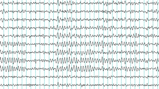
The electroencephalogram allows us to measure, as we have mentioned, electrical activity of the brain Regardless of the objective of the encephalogram, said activity can occur in the form of various types of waves.
Measurements can be taken while awake or during sleep, depending on the purpose for which the test is being done. Through the electrodes, the measurement system captures the emission of brain waves and their rhythm, shape, duration and frequency of emission.
Types of waves
The waves captured They can be alpha, beta, theta and delta Each one will cause the electroencephalograph to draw one or another pattern of wave frequencies.
alpha waves appear in moments of relaxation or when faced with tasks that do not require concentration or effort.
Beta waves are usually reflect the performance of intense mental effort generally appearing while we are awake or during REM sleep.
Theta waves are observed just like alpha waves when we are relaxed, but in this case They are more frequent at times when, in addition to being relaxed, we are sleepy being the most predominant wave type during phase two of non-REM sleep.
Lastly, delta waves are those linked to deep sleep being those that have traditionally been linked to rest and repair of nervous tissues.
Through the encephalogram, both the general functioning pattern of the brain and the differences between some areas with others can be measured, through the analysis of voltage differences between different areas.
Test performance
The basic operation of this technique is not very complex. The test is based on the placement of a series of electrodes at strategic points on the head attaching to a small cloth helmet previously placed on the scalp of the patient or study subject or directly on the scalp.
The record used measures a voltage difference between two electrodes these being placed in pairs to be able to carry out measurements.
Phases of use of the encephalograph
First of all, we proceed to prepare the test, seating the subject to be evaluated and fixing the elements that allow brain activity to be recorded. For it A type of hair gel is applied that improves the conduction of electricity and fix the electrodes more precisely, the collation of which is done below. Generally, around twenty electrodes are placed, creating a setup that allows obtaining a correct view of the activity of the nervous system.
In this setup, it is common to use the 10/20 system, placing the electrodes equidistantly so that they are separated between 10 and 20% of the brain axes. Furthermore, the montage can be bipolar, if the aim is to record brain activity and the difference between two points, or monopolar if a specific point is compared with one without brain activity.
Once the electrodes are placed, the measurement is carried out, first recording the individual’s basal rhythm both with eyes closed and open, and then causing light stimulation to observe the reaction of brain activity. Some common stimuli are slight photostimulation or hyperventilation of the patient. The subject may also be asked to do some type of physical or mental activity.
As the test is performed, a series of results are obtained that indicate how the nervous system acts and how it reacts to stimulation.
The results obtained by the measurement They can be recorded and either printed or directly reflected on a monitor But the recording of the waves does not have a significance in itself, and an analysis of the implications of the basal functioning and/or any alteration detected over the time that the recording has occurred must be carried out.
Uses and applications of the electroencephalogram
Considering all of the above, it must be taken into account that the use of the electroencephalogram is not done on a mere whim. It is used only for specific purposes and when certain ailments are suspected or an investigation is being carried out.
Regarding research, the electroencephalogram is used in those experiments in which it is required to know the brain activity in a certain state or while carrying out specific actions. Thus, it serves to explore how our brain works and how it reacts to specific stimuli or activities. It also allows you to evaluate whether there are large differences between the activation of a specific area and others.
Regarding its use in medicine, can be used to detect whether the brain is functioning normally control the state of consciousness during a surgical intervention or if there are alterations in the wave emission pattern.
In this aspect, this type of technique tends to be used when the presence of disorders such as epilepsy is suspected (even voluntarily provoking seizures to record how and what happens), dementia, encephalopathies, typical outbreaks of some mental disorders and even differentiate between coma and brain death (while in the first there is brain activity, the second would show a flat electroencephalogram). It is also widely used to analyze sleep problems and disorders.
Contraindications and adverse effects
The application of an electroencephalogram does not usually cause problems in those in whom it is performed, being a non-invasive technique which does not present contraindications in the majority of the population, not even in pregnant women.
One of the few exceptions are cases of epilepsy in which a seizure could occur during the test, which in many cases is sought in order to identify hyperactivated areas. However, in serious cases the risk of causing a new crisis would have to be assessed.