Corticospinal Pathway: Characteristics And Functions
The corticospinal pathway is the main voluntary motor control system of the central nervous system
Its axons travel from the cerebral cortex to the spinal cord, and are partly responsible for allowing us to move our limbs and trunk, and for carrying out, together with other nervous tracts, finer and more precise movements.
In this article we explain what the corticospinal pathway is, what its main characteristics are and the functions it performs, as well as the clinical signs and symptoms that occur due to injury to this nervous tract.
The corticospinal pathway: definition and characteristics
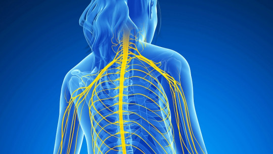
The central nervous system is a complex network of components that allow an organism to interact with its environment It is made up of multiple parts that perform different functions. The upper motor neurons are located in the cerebral cortex, which send movement signals to the lower motor neurons that tell the muscles whether they should contract or relax.
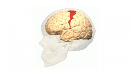
The corticospinal pathway It is made up of the axons of motor neurons that travel from the motor cortices (primary motor cortex, supplementary motor area and premotor cortex) to the spinal cord. These neurons control voluntary movements of the limbs and trunk. Small nuclei of neurons also originate in areas of the parietal lobe (ascending parietal gyrus and superior parietal cortex).
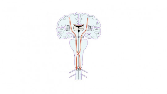
This motor system is one of the last to develop, since the fibers of the corticospinal pathway finish myelinating approximately 2 years after birth. One of the characteristic aspects of this axon bundle is the so-called pyramidal decussation : This means that a large part of the corticospinal fibers (around 75-80%) cross to the contralateral side of the medulla oblongata, and the nerve fibers from the left side of the brain pass to the right hemibody, and vice versa.
Pyramidal decussation leads to an obvious conclusion, which is that the areas of the brain that control the right part of the body are located in the left hemisphere, and those that control the left part are in the right hemisphere. This can be verified when an injury occurs in one of the hemispheres; For example, some patients who have suffered damage to the left hemisphere may suffer from paralysis on the right side of their body.
Neuroanatomical organization
The corticospinal pathway originates in various areas of the cerebral cortex , mainly in the primary motor cortex (Brodmann area 4) and in premotor areas (Brodmann area 6). However, they can also originate in the somatosensory cortex, cingulate gyrus, and parietal lobe. This pathway connects these brain areas with the gray matter of the spinal cord.
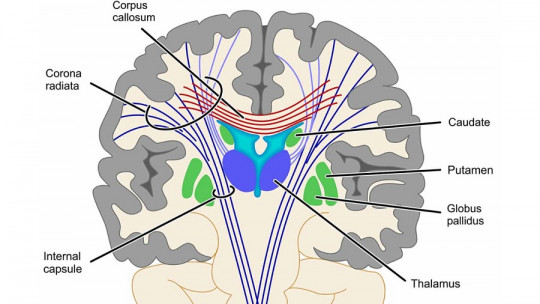
The axon bundles of the corticospinal tract travel from the cortex, through the deep white matter, to the brain stem. The majority of them decussate back and forth in the lower brainstem and descend into the contralateral white matter of the cord in what is called the lateral corticospinal pathway.
About 15% of axons do not perform pyramidal decussation and descend as the ventral corticospinal tract. In addition to the corticospinal pathway, this system contains indirect pathways that project first to the motor nuclei of the brainstem, and from there to the spinal cord.
The gray matter of the spinal cord is the target of this axon bundle. Corticospinal projections from the primary motor and premotor cortical areas are directed to the spinal motor regions, which are composed of the deeper laminae of the dorsal horn, the intermediate zone, and the dorsal horn. The corticospinal system also projects from the somatosensory cortex to sensory processing centers in the dorsal horn and brainstem to regulate proprioceptive information generated during movement.
The corticospinal pathway fulfills an essential function in controlling movements of the limbs and trunk , both in the skill and precision to carry them out. It is also important in the execution of finer movements (such as the fingers), although, in that case, it needs other fibers for its initiation.
It has been suggested that the corticospinal tract is also responsible for modulating the organism’s sensory information, due to the connections it has with the somatosensory cortex. As we have already mentioned, the decussation of the fibers that cross the midline implies that each cerebral hemisphere performs the function of controlling the muscles of the extremities on the opposite side of the body, which does not occur with the muscles of the trunk.
The corticospinal pathway contains pyramidal neurons (Betz cells), from which large axons arise that innervate, mainly, the legs; The special characteristics of this type of neurons allow them to conduct nerve impulses at high speed
Lesions in this part of the nervous system
Lesions in the corticospinal pathway produce a series of clinical signs and symptoms that make up the so-called pyramidal syndrome. Next, let’s see what they are.
1. Impairment of voluntary movements
A characteristic effect of injury to the corticospinal pathway is muscle weakness , whether total (plegia) or partial (paresis), as well as the clumsiness of fine movements of the hemibody on the same side in which the spinal cord damage occurs. This weakness mainly affects the extensor muscles of the upper limbs and the flexor muscles of the lower limbs.
It is common that, after injury to this nervous tract, there is an increase in muscle tone or hypertonia, as well as spasticity in the extremities because the fibers of the corticoreticular pathway that descends together with the pyramidal tract are usually affected.
3. Presence of pathological reflexes
Lesions of the corticospinal tract can cause the presence of pathological reflexes, which are those that can only be elicited in abnormal conditions, which implies an alteration of the central nervous system (e.g. Babinski sign).
4. Increased deep reflexes
Another clinical sign that causes injury to the corticospinal fibers is an increase in deep reflexes. If the corticoreticular fibers are affected, hyperreflexia can occur an increase in the area in which the reflex occurs if triggered by percussion beyond the provocation zone.
There may also be a diffusion of reflexes if the response affects other muscles, apart from the one corresponding to the tendon that has been hit, or a more violent movement if the response is multiple.
5. Other signs and symptoms
For example, the absence of superficial reflexes and muscle atrophy The latter is generally mild, and usually occurs due to a lack of use of the muscle due to motor weakness.