Since the belief appeared that glial cells only exist to provide structural support to neurons it is increasingly discovered that these microscopic elements are closely involved in the proper functioning of the nervous system. Among the usual functions carried out by glia we find defense against damage and invaders, nutrition of neurons or improvement of the electrical impulse, which means that they are much more than a simple support in the development of neurons as they are. as was thought in the past.
From the growing study on glia, we also seek to see how these cells (which represent most of the components of the brain) are involved in neurological diseases and disorders something that until now was only done in research on different types of neurons.
It is important to understand to what extent neuroglia intervenes in these processes, since this may be one of the paths towards finding cures in the future.
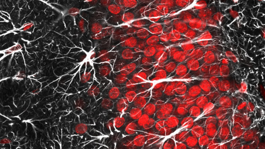
In the Central Nervous System (CNS) we find three main classes of glial cells : oligodendrocytes, responsible for placing the myelin sheath on neurons; microglia, whose function is to protect the brain; and astrocytes, which have a multitude of functions to help neurons.
Unlike the CNS, Only one main type of neuroglia is found in the Peripheral Nervous System (PNS), Sch cells.Wanna which are further subdivided into three. Mainly, they are responsible for generating the myelin layer in the axons of neurons.
Diseases and disorders associated with glia
Currently, There is increasing evidence that neuroglia play a role in diseases that affect the CNS. both for good and for bad. Here I present a small list of them, covering different types of diseases, where I comment on the involvement (which is known today) of glial cells in them. Many more details are likely to be revealed in the future.
Paralysis occurs when the connection between a group of neurons is lost. because their “communication route” has been broken. In principle, glia can release substances known as neurotrophic agents that promote neuronal growth. As occurs in the PNS, this allows mobility to be recovered over time. But this is not the case in the CNS, suffering permanent paralysis.
To demonstrate that glia are involved in non-recovery, since it is the only thing in which this neurological alteration differs when it occurs in the PNS or the CNS, Albert J. Aguayo, carried out an experiment in the 1980s in which rats with damaged spinal cord (i.e. paralysis) received a transplant of sciatic nerve tissue towards the affected area. The result is that in two months the rats were moving again completely naturally.
In subsequent investigations, it has been found that there is a combination of factors that does not allow the total recovery of the connection. One of them is the myelin itself that they produce. oligodendrocytes, which by forming the sheath, prevent the growth of the neuron. The objective of this process is unknown at the moment. Another factor is the excess damage generated by microglia, since the substances it releases to defend the system are also harmful to neurons.
2. Creutzfeldt-Jakob disease
This neurodegenerative disease is caused by the infection of a prion, which is an abnormal protein that has gained autonomy. Another name it receives is spongiform encephalopathy, since the brain of those affected ends up full of holes. giving the sensation of a sponge. One of its variants caused a health alert in the nineties, known as mad cow disease.
Transmitted if ingested, the prion has the ability to cross the selective blood-brain barrier and lodge in the brain. In the CNS, it infects neurons, astrocytes and microglia, replicating and killing cells and creating more and more prions.
I have not forgotten the oligodendrocytes, and it seems that This type of glia resists infection by prions, but does not withstand oxidative damage that appear as part of the fight carried out by microglia in an attempt to defend the neurons. In 2005, it was reported that the normal protein generated by the prion is found in the myelin of the CNS, although its function in it was unknown.
ALS is a degenerative disease that affects motor neurons which little by little lose functionality, causing loss of mobility until reaching paralysis.
The cause is a mutation in the gene that encodes the enzyme Super Oxide Dismutase 1 (SOD1), which carries out a fundamental function for the survival of cells, which is the elimination of oxygen free radicals. The danger of radicals is that they unbalance the charge in the cytoplasm, which ultimately leads to cellular malfunctions and death.
In an experiment with mice with a mutated variant of the SOD1 gene, it was seen how they develop the ALS disease. If the mutation in the motor neurons was prevented, the mice remained healthy. The surprise appeared with the control group, where only the motor neurons showed the mutation. The theory indicates that in these mice the motor neurons would die and generate the disease. But this did not happen, and to everyone’s surprise, the mice were apparently healthy. The conclusion is that the cells close to the motor neurons (glia) had some mechanism associated with SOD1 that prevents neurodegeneration.
Specifically, the lifesavers of the neurons were the astrocytes. If healthy motoneurons cultured in plates joined with SOD1-deficient astrocytes, they died. The conclusion drawn is that the mutated astrocytes release some kind of toxic substance for the motor neurons, explaining why only this type of neurons die in the development of the disease. Of course, the toxic agent still remains a mystery and the subject of research.
4. Chronic pain
Chronic pain is a disorder in which you permanently the pain cells remain active, without any damage that causes their stimulation. Chronic pain develops when there has been a change in the CNS pain circuit after an injury or illness.
Linda Watkins, a pain researcher at the University of Colorado, suspected that microglia may be involved in chronic pain by being able to release cytokines, a substance that is secreted in an inflammatory response and activates pain.
To check if he was right, he performed a test on rats with chronic pain caused by damage to the spine. He administered minocycline to these, which targets microglia, preventing their activation and, as a consequence, they do not release cytokines. The result was immediate, and the rats stopped suffering from pain.
The same study group found the mechanism by which microglia recognize when an area is damaged. Damaged neurons release a substance known as fractalkine, that microglia recognize and defend by secreting cytokines. The problem with chronic pain is that for some reason, microglia do not stop releasing cytokines, constantly stimulating the production of the sensation of pain, even though there is no longer any damage.
5. Alzheimer’s
Alzheimer’s is a disease that destroys neurons and their communication, generating memory loss. A mark of this disease on the anatomy of the brain is the appearance of senile plaques in different regions of the brain. These plaques are an aggregate of a protein called beta-amyloid, which is toxic to neurons.
What generates this toxic accumulation is the astrocytes. This type of glia has the capacity to generate the beta-amyloid peptide, since it can process its precursor, the Amyloid Precursor Protein (APP). The reason for this is still unclear.
Another mark is that around the plates A large number of microglia are observed, which in an attempt to defend the tissue, group together to fight against the accumulation of beta-amyloid and releases toxic substances (such as cytokines, chemokines or reactive oxygen), which instead of helping, promote the death of neurons, since it is toxic to them. Furthermore, they have no effect on senile plaque.