Uncinate Fasciculus: Characteristics, Parts And Functions In The Brain
The uncinate fasciculus is a brain tract that is related to the limbic system although today it is unknown exactly what function it has.
What is known is that if it is damaged it can cause various psychiatric problems and memory problems, as well as changes in personality.
It is one of the structures that takes the longest to fully develop and, below, we will discover in more depth what its anatomical trajectory is and what symptoms are related to its injury.
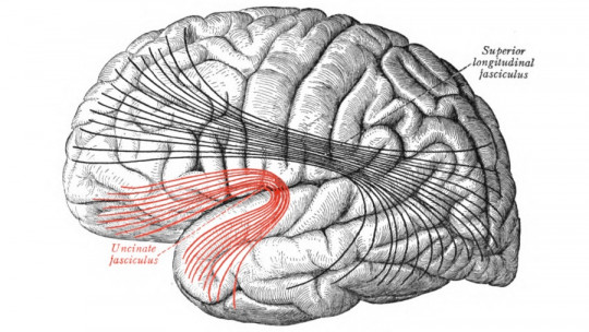
The uncinate fasciculus, classically called the frontotemporal fasciculus, is a white matter association tract in the human brain, which connects different parts of the limbic system such as the parahippocampus, the amygdala in the temporal lobe, portions of the frontal lobe, and the orbitofrontal cortex. It receives its name from its hook shape, and should not be confused with the uncinate fasciculus of the cerebellum or Russell’s tract.
It is not known exactly what its function is, but it is known that It has been seen that its affectation would be behind several psychiatric conditions, such as mood disorders, memory failures and disorders such as schizophrenia. It is known to be one of the last tracts of the human brain to mature, with full maturity being achieved in late young adulthood.
Route and parts
The uncinate fasciculus extends from the basal aspect of the frontal lobe to the lateral aspect of the temporal pole, passing through the M1 portion of the middle cerebral artery in the limen of the insula. This fascicle is made up of fibers that join at the end of the superior, middle and inferior temporal gyri with the fronto-orbital cortex. The fibers also connect to the cortical nuclei of the amygdala and the hippocampus to the gyrus rectus and septal area.
The uncinate fasciculus It can be divided into three segments: temporal, insular and frontal The temporal segment arises from the nuclei of the amygdala, specifically in areas 28, 34 and 36, the perhinal area of the mesocortex (area 35) and the anterior part of the three temporal gyri (areas 20 and 38), linking with the frontal segment in the area of the subcallosal gyrus (area 25), the straight gyrus (area 11) and the posterior orbital cortex in its areas 47, 13 and 14
The fascicle It is a solid tract of fibers between 3 and 7 mm wide and between 2 and 5 mm high, which travels along the lateral part of the extreme and external capsule on the ventral circumference of the putamen towards the retroorbital cortex. The frontal part is oriented horizontally in the straight gyrus, retro-orbital cortex and the subcallosal area.
Within the uncinate fasciculus we can distinguish two types of fibers, there being dorsal or lateral fibers, which occupy the most external part of the tract and which are easier to dissect and separate, and other medial or ventral fibers, more compact and united. Dorsolateral fibers are linked to the pole of the first and second temporal gyri in the lateral retroorbital cortex. The ventromedial fibers connect the uncus, the cortical nuclei of the amygdala, and the tip of the third temporal gyrus to the gyrus rectus and the subcallosal area.
Near the uncinate fasciculus are the capsules or strata of white matter that separate different nuclear formations from the gray one. The claustrum is separated from the striatum by the external capsule, and is related laterally to the cortex of the insula, which are separated by the extreme capsule. The fibers that circulate through these two capsules have a longitudinal path, and interconnect various parts of the cortex with each other. At some specific points in the segments, some fibers of the uncinate fasciculus can lodge inside these capsules.
The medial uncinate fibers filter through the external capsule, while the more lateral filaments pass through the extreme capsule. The most lateral part of the fascicle is part of the extreme capsule and the most medial portion of the external capsule. The cloister is found as if it were a sheet of gray matter between both capsules.
Function
The exact function of the uncinate fasciculus is not yet known, although it is traditionally considered part of the limbic system. It has been proposed that this fascicle allows mnemonic representations to be stored in the temporal lobe, in addition to guiding decision making in the frontal lobe.
Using diffusion tensor imaging, it has been found that this structure has greater activity on the left side than on the right. This has been linked to left hemisphere language specialization. In any case, the use of electrical stimulation in the brain, specifically above the uncinate fasciculus, does not hinder the ability to communicate, which raises doubts as to whether it has any relationship with language.
What is believed is that could play a role in some types of learning and memory, although not in all of them. It seems to be especially involved in learning through stimulus-reward. It has also been related to name-object/person learning, since lesions in this region imply deficits in the memory of names.
Development
The uncinate fasciculus It is one of the regions of the brain that takes the longest to complete its development, reaching full maturity at age 30. Problems in remembering names, reward learning and having impulsive decision making have developed with having a poorly developed uncinate fasciculus.
It is a very vulnerable region. Abnormalities in the left anterior uncinate fasciculus have been found in 12-year-old premature boys. Fractional anisotropy has been observed in 10-year-old children with socioemotional deprivation of the left uncinate fasciculus reduced compared to that of another child, which is related to behavioral, cognitive and socio-emotional problems.
Clinical importance
Malformations and poor development in the uncinate fasciculus have been linked to several neuropsychiatric disorders, including social anxiety, depression and schizophrenia It has also been linked to dementias, such as Alzheimer’s disease, semantic dementia, and temporal lobe epilepsy.
It has also been related to psychopathy and violent behavior, something seen in a 2009 investigation in which damage to the uncinate fasciculus was related to higher scores on the Psychopathy Checklist.
One of the most famous cases of brain damage in the history of neurology is Phineas Gage This man was a railroad worker, who had an accident in which a steel bar pierced his left frontal lobe. Probably, in this accident his uncinate fasciculus was destroyed, along with other regions. This caused him to suffer personality and behavioral changes, becoming an impulsive person who made bad decisions and did not follow social norms.
I’m Emily Williams Jones, a psychologist specializing in mental health with a focus on cognitive-behavioral therapy (CBT) and mindfulness. With a Ph.D. in psychology, my career has spanned research, clinical practice and private counseling. I’m dedicated to helping individuals overcome anxiety, depression and trauma by offering a personalized, evidence-based approach that combines the latest research with compassionate care.


 Components and Functions")


: Definition, Location and Functions")


