What Is The PreBötzinger Complex? Anatomy And Functions
As a general rule, at rest an adult human being breathes at a rate of between twelve and eighteen breaths per minute. Breathing is fundamental for our survival, a process that we carry out semi-consciously continuously throughout our lives.
But who is in charge of us doing it? What part of our body causes us to carry out this basic function? The answer is found in the medulla oblongata, specifically in the pre-Bötzinger complex
The pre-Bötzinger complex: description and basic location
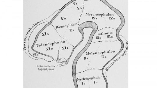
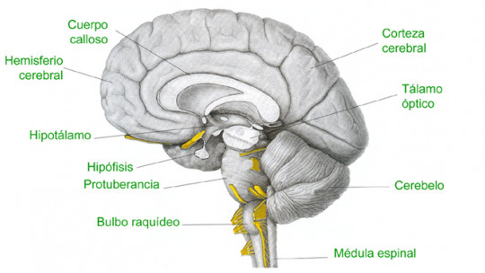
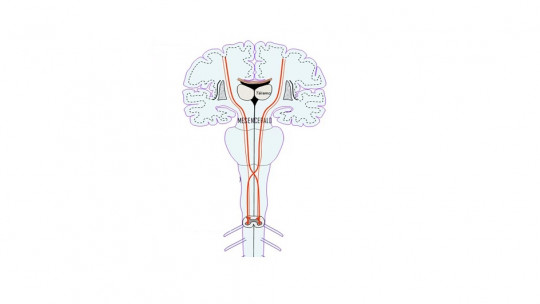
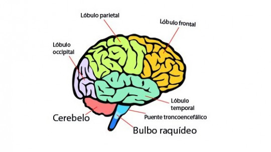
The preBötzinger complex is a set or network of neurons located in the medulla oblongata or medulla oblongata , specifically in its ventromedial part, forming part of the brain stem. This neural network appears in both hemispheres, being a bilateral and symmetrical structure. It connects with the spinal cord, and is, as we have mentioned, essential for the generation and maintenance of the respiratory rhythm.
It is a structure located recently, specifically in 1991, and different types of neurons have been found in it that allow, through their interaction, the genesis and rhythmicity of the respiratory cycle. The preBötzinger complexes of both hemispheres seem to function partially independently, although they communicate in order to synchronize.
Principal functions
Although this structure is still little known, various functions of great importance are attributed to it
1. Basic respiratory rhythm
The preBötzinger complex is a fundamental element to keep us alive, and its injury can cause death due to respiratory depression. Its main function is the generation and management of the respiratory rhythm
Interaction with other areas of the brain means that the preBötzinger complex can regulate breathing rate based on environmental needs For example, if we play sports our breathing will accelerate.
3. Capture of oxygen level
It has been detected that this complex and its connections are capable of detecting and acting based on the oxygen level in the body. For example, If we are suffocating, it is common for our breathing rate to accelerate since the organism seeks to acquire the oxygen necessary to survive.
An unknown mechanism of action
The way in which this structure works is not yet completely clear, but through experiments with rodents it has been shown that it is linked to the hormone neurokinin-1 receptor and to the action of neurotransmitters.
The existence of “pacemaker” neurons has been observed (similar to what happens with the heart rate), some dependent on voltage and others independent of it. Its exact functioning is still debated, although it is speculated that the voltage-dependent ones are the most linked to the generation of the respiratory rhythm by allowing the emission of action potentials through sodium uptake.
In any case The hypothesis with the greatest empirical support is the one that indicates that it is the action of the set of neurons and their interaction that allows the rhythm to be generated being the result of the interaction and not of the activity of a single type of neurons.
Much more research is necessary in this regard to be able to know the exact functioning of this region, being a field of study to be studied in depth.
Regarding the neurotransmitters with the greatest effect in this area, it has been perceived that it is essential that there is glutamatergic activity for the pre-Bötzinger complex to act allowing respiration. Specifically, it is the activity of AMPA receptors that has the most prominence, although some participation of NMDA receptors in the process is also observed (despite the fact that in some studies the modification of NMDA did not generate real changes and do not seem to result essential). Its inhibition can cause the cessation of the respiratory rate, while the use of agonists causes an increase in it
When it comes to reducing the respiratory rate, the neurotransmitters that seem to act the most are GABA and glycine.
In addition to the above, there are other neurotransmitters that influence the respiratory rate through this structure. Although they do not participate directly in the genesis of the respiratory rhythm, they do modulate it. Examples of this are found in serotonin, adenosine triphosphate or ATP, substance P, somatostatin, norepinephrine, opioids and acetylcholine. This is why many substances and drugs cause an alteration in the respiratory rhythm.
One aspect to keep in mind is that emotions also have an important effect on the respiratory rate, due to the effect of the secreted neurotransmitters on this area. For example, in the case of experiencing nervousness or anxiety, an increase in respiratory rate is observed, while when faced with hopelessness and depression, it tends to slow down.
Effects of injury to this area
Although the preBötzinger complex is not the only element involved in respiratory control, it is currently considered the main element responsible for governing it. Alterations in this area can cause consequences of different magnitude, such as respiratory increase or depression And this can come from congenital injuries, trauma, cardiovascular accidents or administration of psychoactive substances. In extreme cases it can lead to the death of the patient.
It has been observed in the postmortem analysis of people with dementia with Lewy bodies or atrophy, a decrease in the population of neurons reactive to the aforementioned neurokinin-1 is usually observed, which may explain the presence of respiratory alterations in these diseases.