Ruffini Corpuscles: What They Are And How These Receptors Work
Ruffini corpuscles They are a type of mechanoreceptors that are especially responsible for temperature perception, which could be considered a subcomponent of the sense of touch.
Although they are very small cells, the truth is that without them we would not be able to conveniently detect when we are in an environment in which we could catch a cold or die from a heat wave, in addition to being important in detecting the stretching of the body.
Neurobiology has been responsible for studying this component of the human sensory system, describing and classifying it in the way it is described in this article. Let’s understand how Ruffini corpuscles are and work below.
Ruffini corpuscles, also called bulbous corpuscles, They are cells which detect sensory stimuli at the skin level, having an important role in constituting and forming the sense of touch. They receive their name from the surname of the person who discovered them, Angelo Ruffini, a notable Italian doctor and biologist.
They are a type of mechanoreceptors that allow detecting changes in temperature and stretching of the skin They have the ability to detect signals within very small receptive fields, which makes them fall into the category of type I mechanoreceptors. They are not very numerous nor are they large in size.
It has been seen that deep skin alterations due to scars, degenerative processes, aging or poor joint disposition can alter the location of these corpuscles.
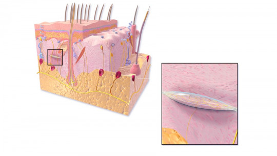
What are these cells like?
Ruffini corpuscles They are made up of many free nerve endings, which have their origin in a myelinated axon and constitute a cylindrical-shaped structure. This structure, which has a capsule appearance, the nerve terminals are organized by anchoring between collagen fibers of connective tissue. The axon demyelinates and later bifurcates into two, before forming branched nerve endings.
Despite this, it should be said that there are some differences between Ruffini corpuscles in hairy skin and those found in glabrous skin. An example of this is in the penis, especially in the foreskin, where the corpuscles originate from a single axon which branches several times before completely demyelinating within the connective tissue capsule.
On the other hand, in the case of skin with hair, the axon takes the shape of a spiral that approaches the hair follicle just below the sebaceous gland, where it branches and loses myelin.
Where are they located?
Ruffini corpuscles are found in both hairy and glabrous skin, that is, skin in which there are no hairs, and also in both the hypodermis and epidermis. They are also found in non-superficial structures, such as the menisci, cruciate and lateral ligaments and joint capsules. These cells can be found in most mammals.
However, despite being found throughout the skin, there are differences in the level at which these corpuscles are found depending on whether there is hair or not. In the case of glabrous surfaces, such as the palms and fingers, soles of the feet, lips, penis and pubis, these cells are found at the level of the reticular layer of the epidermis.
Although in the case of structures in which there is hair, Ruffini corpuscles are also found in the reticular layer of the epidermis, between hair and hair, in addition to being located in the connective tissue capsule that covers the hair part. which is inserted at a certain depth into the skin. The group formed by this type of cell and the capsule is called the pilo-Ruffini complex
In the animal world, in addition to the areas we have mentioned, these corpuscles are found in somewhat peculiar places. In the case of some primates, they have been found associated with regions of the dermis close to the hairs found in the nasal mucosa. In birds and some mammals it has been seen that Ruffini cells are found in joints, but only in the fibrous part and in the ligaments.
What function do they play?
The main function of Ruffini corpuscles is the perception of temperature changes, in addition to stretching the skin. Also they can perceive the continued deformation of the skin and inner tissues
These structures are of vital importance since they are what allow temperature variations to be detected, especially taking the body’s own temperature as a reference, thus establishing whether the environment is colder or warmer and how pleasant it is. They are also capable of detecting mechanical deformation of the skin, although this function is more typical of other mechanoreceptors, such as the Pacinian corpuscles.
In fact, they differ from this other type of skin receptors by the fact that Ruffini corpuscles are slow to adapt. This means that They are able to detect stimuli held on the skin in addition to the slight stretches that can be exerted on this tissue.
It is worth highlighting the fact that they are not only able to detect the stretch, but also perceive the joint angle, the speed of the mechanical stimulus on the skin and the type of stretch.
General aspects of mechanoreceptors
In the sense of touch they have prominence up to four different types of mechanoreceptors One of them is the Ruffini corpuscle, in addition to those of Pacini, Merkel and Meissner.
They all have in common that they are found in the skin, and they respond to physical changes that can occur on this tissue. They act as if they were signal transducer systems, converting mechanical stimulation into electrochemical stimulation, being sent to the central nervous system to be able to organize a response if necessary.
Signals are sent in the form of burst nerve discharges and depending on the characteristics of the sensory cell itself such as the type of stimulus it is responsible for, the stimulation will be continuous or, on the other hand, it will progressively decrease.
These types of cells have been classified based on their behavior during the course of two phases: dynamic and static Dynamic phase refers to the moment in which the intensity of the stimulus varies, for example, when heat is applied and stopped being applied to the skin. On the other hand, the static phase is understood as the moment in which the stimulus does not change its stimulation intensity on the organism.
Those receptors that are only stimulated during the dynamic phase have been called rapidly adapting or phasic mechanoreceptors and this is the case of Pacinian corpuscles.
On the other hand, those that are stimulated during both the dynamic and static phases are known as slowly adapting mechanoreceptors being the case of Ruffini’s.
On the other hand, there is a second classification, depending on the size of the area that these types of receivers are in charge of. Type I receptors are those that receive signals or are responsible for the stimulation of small receptive fields, while type II receptors are responsible for larger receptive fields.