Our brain is made up of a large number of neurons that fit together like an immense puzzle. Thanks to the fact that all of them are in their correct position, our nervous system can function at full capacity and without any type of problem.
However, the neurons are no longer born in their final position. Rather, they are formed in another region of the nervous system and must travel a long path to reach their destination. This phase of brain formation is known as neuronal migration Any anomaly in its development can cause serious malformations in our nervous system and, as a consequence, a large number of neurological disorders.
Our brain is made up of hundreds of thousands of neurons. A large number of these nerve cells they originate in different locations than those they will occupy once adulthood has arrived
This process is known as neuronal migration, and most of it occurs during embryonic development, specifically between 12 and 20 weeks of gestation. During this period, neurons are generated and travel through our brain until they settle into their final position.
This movement is possible thanks to signals from other neurons, which are already in their final position and play a role similar to that of a traffic light that directs traffic, sending different types of signals to which the neurons in the process of movement respond. migration.
This migratory procedure occurs from the ventricular zone of the neural tube, the place where neurons originate, to the place designated for them. During the beginning of neuronal migration, these cells They are located between the ventricular zone and the marginal zone which form the intermediate zone, a space of transitory location.
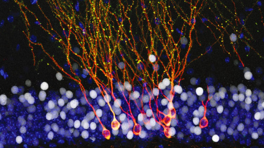
Neuronal migration takes place in different phases and is highly complicated. since these nerve cells must travel a long distance and avoid numerous obstacles so that the brain can develop completely and satisfactorily. For it, They are helped by a type of cells that form what is known as radial glia and which acts as a scaffold through which migrating neurons move.
When some of these phases of neuronal migration are not carried out correctly, changes in the organization of the brain can appear, up to very important brain malformations.
Migration phases
As mentioned in the previous section, the neuronal migration process occurs in different phases, specifically in three, of which each and every one of them is essential for successful cortical formation. These stages of neuronal migration are as follows.
1. Cell proliferation phase
In this first phase, which occurs from day 32 of the gestational cycle, nerve cells or neurons originate.
A large number of these neurons are born in the germinal zones or germinal matrices, hence the name of the phase. These areas are located in the walls of the lateral ventricles.
2. Neuronal migration phase
Throughout this second phase, neuronal migration itself occurs. That is, the neurons leave their place of origin to head towards their final position.
This process occurs thanks to the radial glial system. In this system, a cell that is no longer present in the adult brain guides neurons into position.
3. Horizontal and vertical organization phase
In this last phase, the differentiation and subsequent organization of neurons takes place. Due to the complexity of this final stage, what it consists of and what its particularities are will be explained below.
When the neuron has managed to reach its final location, the differentiation phase begins, achieving all the morphological and physiological qualities of a fully developed neuron. This differentiation depends both on how said neuron is genetically preconfigured, as well as on the interaction with other neurons and the creation of connection pathways.
In our nervous system, as in the rest of vertebrates, neural cells differentiate from each other as a result of different progenitor cells; which are located in specific locations of the neural tube.
Once the differentiation process is finished, neurons organize by joining each other ending the neuronal migration process and completely ending the development of our brain.
Defects in this biological process
As detailed in the first point, any abnormality in the course of neuronal migration can have consequences on the formation of our brain ; from malformations to alterations in brain organization.
The most serious malformations are associated with alterations in intellectual development and epilepsy, while in organizational problems the brain has a correct external appearance but neural connections are severely damaged because its correct disposition in the brain did not occur.
Among the causes of these failures are:
Regarding the consequences of these defects on migration. An abnormal development of the process can give rise to a large number of disorders and disorders. Among these disorders we can find:
1. Lissencephaly
Lissencephaly is the most serious consequence of a failure in neuronal migration. In this case, the neurons begin their migration but are not able to complete it, which causes serious deformities in the brain.
Depending on the severity of the malformation, lissencephaly can be divided into three different subtypes:
2. Periventricular heterotopia
In this case, the problem is due to an alteration in the beginning of the migration. This affects a small group of neurons which accumulate in locations different from those to which they normally belong.
In these cases, the person experiences severe seizures that emerge during adolescence Furthermore, although they usually have normal intelligence, certain patients experience learning problems.
3. Polymicrogyria
In polymicrogyria, the arrangement of the neural mass creates small abnormal gyri that are separated by superficial sulci, creating an irregular cortical surface.
In this condition, two types of polymicrogyria can be distinguished with different clinical pictures:
4. Schissencephaly
Schisencephalies are distinguished by presenting a normal volume of gray matter but with alterations in the gyri that are smaller and more superficial than usual and surrounded by very shallow sulci.
This pathology does not have specific clinical symptoms, but these may vary depending on the extent and location of the affected areas. In some cases there may be no visible clinical symptoms, while in others people may suffer epileptic episodes of varying intensity.
5. Others
Other neurological alterations that are caused by an alteration in neuronal migration are: