Embryology is the science that studies the development of a new human being. This covers from fertilization to birth, although some books also include the formation of gametes called gametogenesis.
It is a complex science that includes the investigation and explanation of all the changes and processes that occur in the formation of a new being. In this article we detail the different stages from the beginning of pregnancy to its end, that is, the stages of embryonic development
In its development, the embryo goes through a series of stages and decisive processes over 40 weeks. Embryology divides these weeks into pre-embryonic period, embryonic period and fetal period.
The embryonic period ranges from fertilization (which occurs on the day established as zero) until the acquisition of a three-dimensional configuration in week 3. In the embryonic period, the outlines of all the baby’s future organs are formed, this goes from week 4 to 8. From week 9 we enter the fetal period where the organs and systems finish growing and acquire all their functions so that birth is possible.
1. Pre-embryonic period
As we have said in the introduction, embryonic development begins with fertilization, this is established as day 0 of the development of the pre-embryo. Fertilization refers to the meeting of a male gamete (sperm) with a female gamete (type two oocyte) in the fallopian tube (tube-shaped structure that connects the ovaries to the uterus).
The pre-embryonic period lasts until the true embryo is formed, that is, when it no longer has a layered or lamellar configuration. The meeting of the gametes produces a single cell called an egg or zygote. The single-celled structure originally found in the ampulla (the upper third of the fallopian tube) begins its journey to the uterus.
1.1. First week of preembryonic development
This week’s goal is to reach the endometrium (the mucosa of the uterus), since this is the most ideal point for the successful implantation of the cellular structure and its growth.
On its journey through the fallopian tubes, the zygote undergoes a cell division process known as segmentation This divides into 2 daughter cells, then into 8… And so on. These cells are known as blastomeres.
Thus, although it grows in number, the mass of cells does not grow in size, since it is initially surrounded by two thin membranes: the internal pellucid membrane and the external corona radiata membrane. This gives rise to a phenomenon known as compaction. The cells acquire a polarity: they are concave on the outside and convex on the inside.
This particular arrangement gives this mass a blackberry appearance, which is now called morula. The morula appears specifically on the third or fourth day of preembryonic development and contains between 16 and 32 cells. It should be noted that the process of segmentation – or cell divisions – is exponential. The first division occurs 24 hours after fertilization; However, the others are considerably reducing this time. An average newborn has 15 billion cells.
The morula and the compaction phenomenon give rise to a cavity that is located in the center of the structure. That’s good, The cellular structure is now hollow and a fluid called blastocoel begins to penetrate This becomes called a blastocyst (immature cavity) that already contains two types of differentiated cells (day 5). The trophoblast, from which the embryonic annexes are formed (amnion, yolk sac, allantois, chorion and placenta). The embryo itself is derived from the outermost layer. The embryoblast produces all human tissues.
When it reaches the endometrium (between day 5 and 6), in order to implant in the mucosa, the blastocyst has to break the membranes that surround it. This process is known as hatching. In summary, at the end of the first week of development we have a spherical structure differentiated into two layers of cells (trophoblast and embryoblast) that has reached the endometrium.
1.2. Second week of pre-embryonic development
In the second week, implantation in the uterine mucosa continues and several changes occur at the intraembryonic level.
First of all, The innermost layer – the embryoblast – is divided into two distinct layers: the epiblast and the hypoblast At this moment we can describe the embryo (remember that it originates from the embryoblast) as a mass of flat cells. This takes the name of bidermal or bilaminar embryonic disc. This first differentiation already allows the establishment of a dorsal (epiblast) and ventral (hypoblast) axis of the embryo.
It is from the epiblast that all the structures and tissues of the body originate. Also, from this, the first embryonic cavity is formed: the amniotic cavity which at some point in development will contain the embryo.
The amniotic cavity originates from an “excavation” of the epiblast cells in contact with the trophoblast. This is quickly covered by flat cells derived from the epiblast known as amnioblasts. The amnioblast is responsible for producing amniotic fluid. A layer of flat cells dissociates from the epiblast. These cells are called amnioblasts and produce amniotic fluid. Finally, it should be noted that this cavity grows progressively.
Cells migrate from the hypoblast into the cavity of the blastocoel to form the primary yolk sac This is called Heusser’s membrane, or exocoelomic membrane. This is a combination of hypoblastic cells and short-lived extracellular matrix.
Meanwhile, the layer of cells that surrounds the sphere, the trophoblast, also divides into two sheets or layers. The syncytiotrophoblast, an undifferentiated tissue that has the mission of invading the uterine mucosa; and the cytotrophoblast, an internal cellular tissue that will serve as an anchor for the embryonic chorion to the maternal endometrium. These two tissues will allow the utero-maternal circulation system to be formed.
At the end of the second week, the pre-embryo is completely implanted in the endometrium of the mother’s uterus. Implantation can cause small bleeding that is sometimes confused with menstruation.
1.3. Third week of preembryonic development
A trilaminar embryonic mass emerges in the third week of development; This process is known as gastrulation This trilaminar germ disc houses three distinct embryonic layers: an ectoderm, a mesoderm, and an endoderm.
Epiblast cells proliferate very quickly, so they begin to migrate and occupy new places. Thus, the epiblast moves and indirectly displaces the cells of the hypoblast, which in turn gives way to two new embryonic layers: the endoderm and the mesoderm. These three layers establish the beginning of all the organs and tissues derived from our body
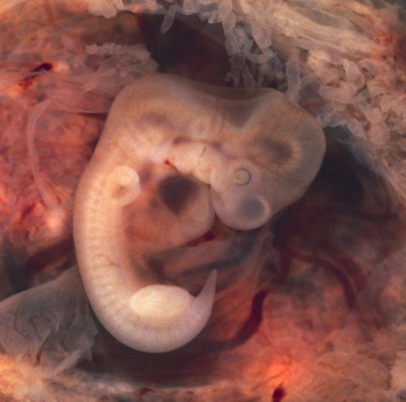
2. Embryonic period (4 to 8 weeks)
The embryonic period occurs between the fourth and eighth week. At that moment, the conceptus or preembryo changes from a flat shape to a cylindrical shape This process is known as folding.
The main biological process that occurs during this stage is organogenesis. During this time, the embryo’s organs begin to develop, eventually leading to the creation of future systems and structures. Embryonic cells proliferate and begin to behave in specific ways. The heart, muscle, gland and future nails draw the first outlines in the embryo.
Of all the systems, the nervous system is the first to appear. This develops from a structure known as the neural tube or epineuria (in reference to its appearance on the outside of the embryo). The process of formation of the nervous system is known as neurulation. It should be noted that the lungs will not be functional until the moment of birth; This means that not all organs evolve in the same way. The heart, for example, already has its structure with the four chambers and the great vessels around week 8.
During this period, the embryo goes through what is considered the most dangerous stage. It is more susceptible to teratogens, or harmful agents, that can cause mutations. Consequently, there are greater chances of developing abnormalities, whether mild or serious.
3. Fetal period (8 weeks at the end)
As we have seen, the changes that occur in the embryo are progressive. However, the transition from name to fetus means that outlines of all the important systems already exist. The growth of the fetus accelerates during this time, and the tissues and organs of the fetus differentiate and specialize in their different functions. Finally, the fetus remains in the uterus during this period, which is known as the fetal period
In the fetal period, the head stops developing faster than the rest of the structures. Additionally, over time, the fetus matures and develops defenses that reduce the chances of miscarriage.
Conclusion
Learning the basics of embryology can help doctors determine the status of a pregnant patient and the developing newborn. As shown in this article, the life cycle begins with the formation of a single-celled embryo and ends with its appearance in the world. Thanks to its findings, this specialty helps families understand possible anomalies before birth and also provides treatments that ensure that the embryo continues to develop normally, without complications.


")


