Neurulation is the process by which the neural tube forms during intrauterine development. The neural tube is essential for the differentiation of the cells of the central nervous system, while the neural crests, structures associated with the one in question, are essential for the formation of the peripheral nervous system.
In this article we will describe the two phases of neurulation or formation of the neural tube: the primary one, in which the neural plate begins to fold back on itself, and the secondary one, which completes this process and allows the subsequent development of the nervous system.
The neural tube is an embryonic structure that forms during the first month of gestation; Specifically, the tube has just closed around week 28 after fertilization. It is the precursor of the central nervous system composed of the brain and spinal cord.
As embryonic development progresses, the neural tube divides into four sections: the forebrain (prosencephalon), the middle brain (midbrain), the posterior brain (hindbrain), and the spinal cord. Each of these parts will progress until giving rise to the different elements that make up the adult central nervous system.
While most of the nervous system develops from the walls of the neural tube, the gap found between the walls is also relevant: the neurocele or neural canal. This structure will progressively transform into the ventricles and the rest of the brain cavities, through which the cerebrospinal fluid circulates.
After fertilization, the zygote is formed, the original cell composed of the fusion of an egg and a sperm. The zygote divides successively, becoming a group of cells called a morula. Later, the blastocoel, a fluid-filled cavity, appears within this structure; When this happens we speak of a “blastula”.
Later The blastula is divided into three layers: the endoderm, the mesoderm and the ectoderm Each of these sections will give rise to different parts of the body. The ectoderm is the most important for the matter at hand, since the nervous system, both central and peripheral, develops from it.
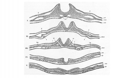
The notochord, a structure located in the mesoderm, sends signals to the cells around it. Those that do not receive these signals become the neural plate or neuroectoderm, a set of cells that have already specialized in nervous functions. The word “plate” refers to the flattened appearance of the neuroectoderm.
Primary neurulation consists of proliferation of nerve cells in the neural plate These cause the plate to transform into the neural tube, a fundamental step in the development of the human body.
Formation and closure of the neural tube
During the neurulation process, the neural plate flattens, elongates and folds on itself around the neural groove, which ends up becoming U-shaped as the walls rise. forming the neural crests and neural tube At this point in the process the tube is open at both ends; we refer to the caudal and rostral neuropores.
Normally these openings close after a few days; however, sometimes the tube does not close properly which gives rise to disorders such as spina bifida (affecting the spinal column) and anencephaly (associated with very serious malformations in the brain).
It is important to differentiate the neural tube from the neural crest because the former transforms into most structures of the central nervous system, while the peripheral is a progression of the neural crest.
secondary neurulation
Secondary neurulation is the process that culminates the formation of the neural tube This is not due to the signals sent by certain cells, as occurs with primary neurulation, but rather occurs as a consequence of the development of the neural tube itself.
This process is associated with the division of neural tube cells between mesenchymal and epithelial. The first are located in the central part of the tube, and the second in its peripheral region. As these cells differentiate, cavities form between the two sets.
The mesenchymal cells that are located in this part of the embryo condense and form what we know as the medullary cord; This, in turn, hollows out inside until it makes way for the cavity of the neural tube. This phenomenon begins in the sacral region of the spine
Thus, while primary neurulation consists of the folding of the neural plate on itself, secondary neurulation corresponds to the emptying of the cavity of the neural tube, closely associated with the differentiation of the cells of the embryo’s nervous system.