Lung Alveoli: Characteristics, Functions And Anatomy
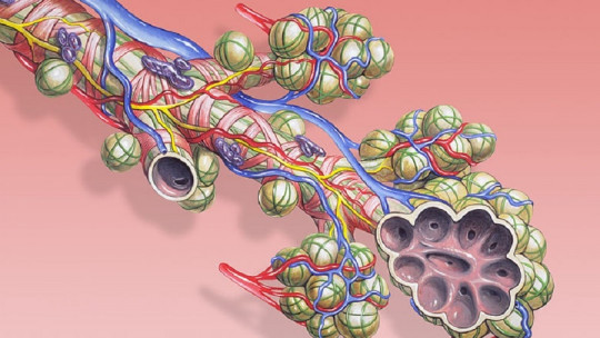
At the most distal point of the bronchial tree there are small structures grouped in the shape of a bunch of grapes that are crucial for our life: the pulmonary alveoli.
In them, the gas exchange of respiration occurs, allowing the entry of oxygen into our body and the expulsion of toxic carbon dioxide, in addition to fulfilling other functions.
Next we are going to see in depth what the pulmonary alveoli are what their anatomy is like, what cells make them up and how they carry out gas exchange.
The lung alveoli are microscopic air sac-like structures found in our lungs, at the ends of other structures, the bronchioles They are often described as shaped like a raspberry or a bunch of grapes. Each alveolus measures approximately 0.2 to 0.5 mm in diameter and is delimited by a wall made up of very thin cells called pneumocytes. On average, an adult person has more than 500 million alveoli which, if stretched, would occupy an area of 80 square meters, the equivalent of a tennis court.
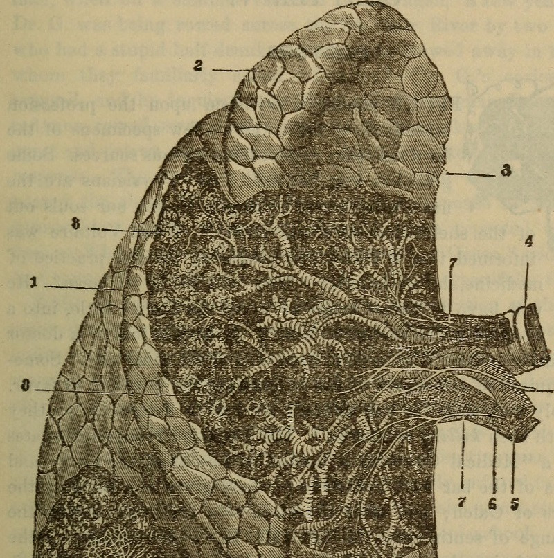
The human respiratory system is made up of several structures, each with specific functions. For example, the conduction system is what allows the passage of air from outside to inside the body and vice versa, being formed by the nasal cavity and passages, the paranasal sinuses, the pharynx, the larynx, the trachea, the bronchi and the bronchioles. The alveoli They are located just at the most distal end of the conduction system specifically at the end of the respiratory bronchioles, grouped in alveolar sacs or acini.
The respiratory functions of the lungs are highly determined by the alveoli, microstructures that represent more than 90% of its total volume and that constitute the lung parenchyma Through the wall of the alveoli, gas exchange takes place between the inspired air and the blood that circulates through the blood capillaries found in the thin walls that give shape to the bronchioles.
Some respiratory diseases cause the alveoli to be seriously affected, such as asthma or tuberculosis, conditions that make the quality of life of the affected person very difficult if they do not receive adequate treatment.
The pulmonary alveoli are found in acini or alveolar sacs, groupings or clusters with a shape similar to a raspberry, a bunch of grapes or a honeycomb. They are defined as the blind-ended units located after a transitional bronchiole, that is, where a terminal bronchiole ends and a respiratory bronchiole begins. Within each acinus, all air conduction pathways or channels have alveoli attached to their walls, participating in both conduction and gas exchange. Approximately, an adult human lung has 30,000 acini.
We can describe the alveoli as sacs with a polyhedral structure that, as mentioned, are between 0.2 to 0.5 mm in diameter. The alveoli are separated from each other by a septum. The air that is introduced into the alveolus of an acinus can be transferred to the other alveoli of the same sac through small pores, since the alveoli that make up an alveolar sac are closely related to each other.
The pulmonary capillaries pass through the septa These ducts are thin branches of the pulmonary arteries, through which blood rich in carbon dioxide (CO2) and poor in oxygen (O2) circulates. The destination of this blood is gas exchange. These septa or alveolar walls are very thin, just 0.5 mm thick, made up of a thin layer of connective tissue that contains extracellular matrix components and different types of cells.
The alveolar walls, better called respiratory membranes, serve as a separation barrier between the air in the alveoli and the blood It is made up of squamous alveolar cells, squamous capillary endothelial cells, and a basement membrane.
Types of alveolar cells
There are three types of cells that we can highlight in the lung alveoli.
Type I pneumocytes
Type I pneumocytes or squamous alveolar cells They are the most abundant cells on the surface of the alveoli, covering approximately 95% of their area They are thin and broad cells, whose thin walls allow rapid diffusion between air and blood, facilitating gas exchange in the alveoli.
type II pneumocytes
Type II pneumocytes or granular pneumocytes They are cuboidal cells, with apical microvilli and have abundant rough endoplasmic reticulum and Golgi apparatus They occupy about 5% of the surface of the alveolus. They do not intervene in gas exchange itself, but they contribute to making breathing possible by facilitating distension and recovery of the size of the alveoli.
Surfactant is composed of phospholipids and proteins that “shelter” both the alveoli and the small bronchioles in order to prevent pressure buildup and alveolar collapse when exhaling. If it were not for the surfactant, the walls of the deflated alveolar sacs could collapse together as if they were sheets of wet paper, making it very difficult to fill them during the next inhalation.
Related article: “Erythrocytes (red blood cells): characteristics and functioning”
Alveolar macrophages
The most numerous lung cells are alveolar macrophages, also known as dust cells These cells slide between the alveolar lumen and the connective tissue, cleaning the surface of any foreign agent through phagocytosis. Its function is to eat particles of dust, pollen or other foreign agents that may have passed through the upper portions of the respiratory tract. If the lungs are infected or hemorrhaged, macrophages are responsible for phagocytosing bacteria and blood cells.
Every day, 100 million alveolar macrophages die as they travel up the alveolar ducts and through the mucociliary staircase, to be swallowed in the esophagus and digested as part of the process of removing dirt from the lungs.
You may be interested: “Macrophages: what they are, characteristics and functions”
Its main functions
The alveoli are the most distal structures of the respiratory system, which makes them carry out functions of vital importance for external respiration Among them we highlight:
They increase the surface area for gas exchange.
They facilitate gas exchange between air and blood.
They expand during inhalation to fill with O2-rich air.
They contract during exhalation to empty CO2-rich air.
Its macrophages protect us from harmful substances, particles and microorganisms.
Next we are going to delve into its main function in the gas exchange process.
Related article: “The 7 parts of the lung: functions and characteristics”
The gas exchange
Respiration is an essential process for most living beings and the cells that make them up. Breathing not only involves introducing enough oxygen into our body to keep it alive and allow various vital functions to continue to be carried out, but It also involves the elimination of waste products produced by the metabolism If they are not eliminated, they can accumulate causing severe damage to the body.
What we know as respiration actually comprises three different but functionally related processes: ventilation, use of oxygen at the cellular level and gas exchange.
Ventilation is the mechanical process that enables the movement of outside air, rich in oxygen, into the lungs ; and the movement of indoor air, rich in carbon dioxide, to the outdoors, expelling it from the lungs.
With the use of oxygen we refer to all the chemical reactions, typical of cellular metabolism, that occur thanks to the presence of this gas and through which the energy necessary for the maintenance of cellular and bodily processes is obtained.
As we have introduced in previous sections, gas exchange is the exchange of oxygen and carbon dioxide between blood and the air contained in the lungs and between blood, organs and tissues.
Specific, The lung alveoli are involved in gas exchange during respiration The air that is introduced into the lungs during inhalation is rich in oxygen, with concentration levels of this gas higher than those in the blood that circulates through the blood capillaries in the alveolar walls. It is thanks to the differences in oxygen concentration between inhaled air and blood that allow O2 to diffuse into our bloodstream.
When the cells of our body receive oxygen from the blood (by diffusion) they use it to obtain energy that can be used to carry out different functions, on which our life depends. This energy comes in various forms, such as ATP and related molecules.
The problem with cellular metabolism, in which oxygen is used, is that some waste is always produced It is not a completely clean process since it produces a waste gas: CO2. The accumulation of carbon dioxide in both cells and tissues is very toxic for our body, so it must be eliminated. Cells get rid of CO2 by releasing it into the blood, where it will be removed from the body during exhalation.
In this way, cells exchange O2 for CO2 with the blood. As this happens, the concentration of the toxic gas increases in the blood, exceeding the level of CO2 concentration in the air. Thus, when blood reaches the alveoli, it exchanges its CO2 for external O2, also causing a change in concentrations.


")

")

