Diencephalon: Structure And Functions Of This Brain Region
When it begins to develop, the central nervous system is made up of three sections: the forebrain, the midbrain, and the hindbrain. Later these initial structures will give rise to the different parts of the adult brain, including the diencephalon.
In this article we will describe the structure and functions of the diencephalon which encompasses regions as important as the thalamus and hypothalamus and allows the correct functioning of multiple biological processes, such as the secretion of hormones and the regulation of the autonomic system.
The diencephalon is a part of the brain that is located in its medial region This name refers to the part of the neural tube that gives rise to various brain structures as embryonic development progresses.
Specifically, once differentiated, the main parts of the diencephalon are the thalamus, hypothalamus, epithalamus, subthalamus and retina Likewise, the pituitary gland or pituitary gland is linked to the hypothalamus, and the optic nerve also connects with the diencephalon.
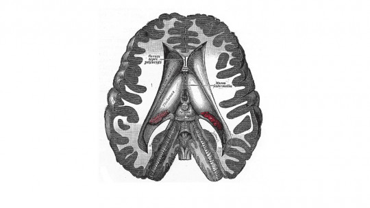
The cavity formed by these structures is the third ventricle, which cushions the effects of trauma that could damage them. The posterior cerebral artery and the circle of Willis provide blood supply to the diencephalon.
Parts of the brain and encephalon
The brain is the part of the central nervous system that is located in the cranial cavity, as opposed to the spinal cord. This organ is made up of the cerebrum, cerebellum, and brainstem
The diencephalon develops from the prosencephalon or forebrain, one of the three parts of the brain at the beginning of the embryonic development of the nervous system. The other two initial sections are the midbrain, which will unite the different parts of the brain, and the hindbrain, which will give rise to the cerebellum, the medulla oblongata and the pons.
As the fetus grows The forebrain divides into the diencephalon and the telencephalon ; From this the cerebral hemispheres, the basal ganglia and the limbic system, including the amygdala, will develop. We describe the sections of the diencephalon in the following section.
Structure and functions of the diencephalon
The brain region that we know as the diencephalon is made up of various structures. These are connected to each other and to the rest of the nervous system, both at the cortical and subcortical levels.
Its relationship with the endocrine system, made up of glands that secrete hormones into the blood, is also very relevant.
1. Thalamus
The thalamus functions as a kind of relay nucleus for connections between the cerebral cortex and subcortical structures It is essential for the reception of sensory inputs (with the exception of olfactory inputs, which go directly to the cortex) and their transmission to the brain lobes.
This structure also plays a role in the regulation of consciousness and the sleep-wake cycle, and influences motor skills through outputs that project from the thalamus to the basal ganglia and the cerebellum.
2. Hypothalamus
The hypothalamus is located below the thalamus. The main functions of this structure include connect the nervous and endocrine systems and control the secretion of hormones by the pituitary gland and other glands.
The hypothalamus directly produces vasopressin and oxytocin, but it also stimulates the endocrine glands to secrete other hormones. It is also key to regulating the body’s homeostasis since it intervenes in thirst, hunger, temperature, circadian rhythms, stress and other bodily processes.
3. Pituitary gland or pituitary gland
The pituitary gland is an endocrine gland attached to the hypothalamus It is very important for growth, kidney regulation, sexual function and reproduction, among other aspects.
It consists of two lobes: the anterior pituitary gland (adenohypophysis) and the posterior pituitary gland (neurohypophysis). While the neurohypophysis secretes oxytocin and vasopressin, synthesized by the hypothalamus, the adenohypophysis produces and releases corticotropin, growth hormone, prolactin, luteinizing hormone and follicle-stimulating hormone, among others.
4. Epithalamus
This brain structure It is mainly composed of the pineal gland, fundamental in circadian and seasonal cycles, and the habenula, involved in the function of the neurotransmitters dopamine, norepinephrine and serotonin. The epithalamus connects the limbic system with other regions of the brain.
5. Subthalamus
The subthalamus is attached to the globus pallidus, one of the main nuclei of the basal ganglia. Due to this, it plays a regulatory role in extrapyramidal and involuntary movements.
6. Retina and optic nerve
The retina develops from the diencephalon, so It is considered a part of the central nervous system The optic nerve allows the transmission of information from the eye to the brain through its union with the diencephalon.
7. Third ventricle
The cerebral ventricles allow the circulation of cerebrospinal fluid, which performs functions similar to those of blood in the brain and spinal cord, in addition to protecting neural tissue from blows and injuries. The third ventricle is located in the middle part of the ventricular system, below the epithalamus.


")


