Cardiac Foci: What They Are, Characteristics And Possible Causes
Heart sounds are the sound expression of the closing of the heart valves. Its operation, at a physiological level, is always unidirectional, which allows blood circulation adequately without organic problems arising. Human beings have about 4.5-5.5 liters of blood in our body and, as surprising as it may seem, the heart is capable of pumping almost all of the fluid in about 60 seconds.
The human heart beats, on average, about 80 times per minute, which translates into 3 billion involuntary contractions throughout our lives. With the size of a fist and the greatest possible resistance, this organ filters about 7,000 liters of blood every 24 hours (or more).
We could continue telling curious facts about the physiology of the heart for hours, because literally, we owe it our lives and our very existence. In any case, today we find it useful to address a more complex and specific topic: stay, then We tell you everything about cardiac foci and, specifically, the intracardiac echogenic focus (EFI).
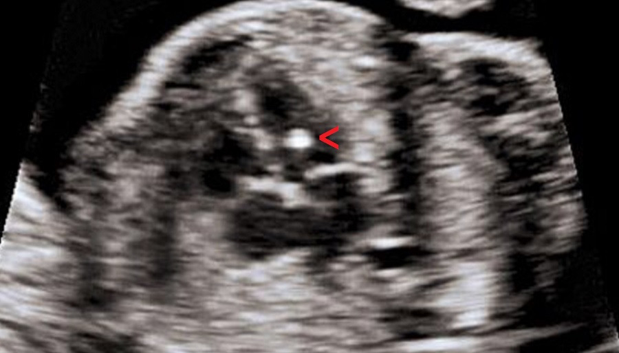
Cardiac foci, more specifically intracardiac echogenic foci, are small bright spots that are seen in the heart of a fetus in an ultrasound examination (ultrasound) We remember that ultrasound uses sound waves to produce images of internal areas of our body, so this technique is very useful to observe the development of the fetus throughout pregnancy.
In the second trimester ultrasound, the structure and function of the heart and fetus are routinely evaluated. In this type of examination, special attention is paid to the 4 chambers of the organ (right atrium, left atrium, left ventricle and right ventricle). As we have said, sometimes small bright “spots” are observed in the heart, generally in the ventricular muscles, which allow blood to be pumped to the entire baby. These are the intracardiac echogenic foci (IEF).
EIF(s) are believed to be normal and harmless in most cases, since they are observed in 5% of fetuses during the second trimester and are not necessarily associated with pathologies at the time of birth or during development. In other words, cardiac foci alone do not endanger fetal health.
Curiously, It has been detected that EIF follows a certain ethnic pattern, since up to 30% of fetuses in Asian people can develop it, while the global average prevalence is 3-5%. It is more common in Asian, African, and Middle Eastern babies, and in almost 80% of cases the ultrasound “flash” occurs in the left ventricle. In 18% of cases it appears on the right, while only 4% of patients experience it on both at the same time.
Intracardiac echogenic foci and their relationship with chromosomal abnormalities
We have said that light bulbs are not bad per se, but science has a lot to argue about this issue. Specifically, research such as Significance of an Echogenic Intracardiac Focus in Fetuses at High and Low Risk for Aneuploidy have shown that, Unfortunately, there is a correlation between FEI and trisomies on chromosomes 21 and 13 We will tell you below what is known about it.
EIF and trisomy 21
Down syndrome is a genetic condition resulting from a chromosomal abnormality, which results in the presence of a more partial or total copy of chromosome 21. Human beings have 2 copies of each chromosome in each of our cells and, therefore , we are diploids (2n). The oddity of trisomy 21, as its name suggests, is that during meiosis chromosome 21 is not distributed well Therefore, the patient ends up with an accessory copy (2+1) and manifests the symptoms of Down syndrome.
90% of cases are due to these meiotic errors, while 4% and the remaining percentage are the product of problems such as balanced translocation and errors after fertilization. In short, this condition causes an extra copy of chromosome 21 and affects 10 in every 10,000 live births.
According to the research cited above, Cardiac foci are present in approximately 18% of fetuses with trisomy 21, compared to 5% of foci experienced in normal babies This could indicate that FEIs could be minor markers for detecting a trisomy, but this correlation is not always true.
However, this does not mean that an EIF has no clinical importance. By itself it is not a trait that should be supported with genetic analysis, but if the mother has certain risk factors, it is time to start performing additional tests.
EIF and trisomy 13
Trisomy 13 follows the same premise as the previous case, that is, the patient has a copy of more than one of the somatic chromosomes, this time number 13. It can manifest completely, partially or in a mosaic, but we It is enough to know that the extra genetic material interferes with the patient’s normal development.
More than 90% of newborn children with trisomy 13 die before their first year of age so we tell a very different story to the previously mentioned disorder.
Things get interesting, on a medical level, when we discover that 39% of fetuses with trisomy 13 present microcalcifications in the papillary muscle (cone-shaped muscular projections, whose bases join the ventricular wall). It is believed that cardiac foci correspond to these formations, that is, the ultrasound detects that there is more calcium than normal in an area of muscle tissue. Naturally calcified tissues, like bones, look brighter on ultrasound, so this correlation makes sense.
As we have said previously, cardiac foci are considered “normal” when they appear occasionally on an ultrasound during 18-20 weeks. In any case, if there is no evidence of pathology, it is classified as isolated events so it is not taken into account when making a diagnosis.
Additionally, many EIFs disappear before the third trimester, but others do not. This scenario is also considered normal, so monitoring for bright spots in cardiac tissue is not followed unless other warning signs appear. Beyond this, other studies cite that the correlation between trisomy 21 and FEI is 1%, so special emphasis is placed on not worrying when this event is found on an ultrasound.
For all these reasons, There are no diagnostic tests for those fetuses that show only isolated cardiac foci Among the possible suspicious pathological events, we find the following:
If none of these criteria are met, the cardiac foci are considered innocuous and no additional tests or monitoring are performed.
Summary
As you have seen, Very little is known about intracardiac echogenic foci, to the point that their causes are not even really known It is stipulated that it is due to characteristic microcalcifications in the heart muscle, but there is not even a clear idea about the etiology of the event.
On the other hand, some investigations associate trisomy 13 or 21 with cardiac foci, while others do not dare to make clear correlations. This physiological event alone does not indicate anything, so it should not alarm the parents of a baby when it occurs in isolation.


")


