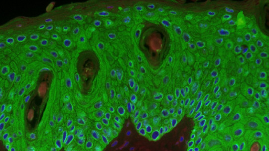
The skin is the largest organ in the human body. With about two square meters of surface and up to five kilograms of total weight, this tissue conglomerate is the most important primary biological barrier of complex living beings, along with the mucosa, saliva, tears, sweat and certain behavioral mechanisms (such as coughing). .
The skin is an unforgiving environment for pathogens, as it is dry, has a slightly acidic pH, has antiseptic properties and, in addition, there are other microorganisms that already colonize this layer without causing us any harm (staphylococci, micrococci and Acinetobacter). All this makes the task very difficult for bacteria and parasites that want to take advantage of us, since they encounter a practically insurmountable physiological and living barrier.
Therefore, it is not surprising to know that the vast majority of skin infections begin with a wound: when a crack opens in this wall, both commensal and pathogenic bacteria take advantage of the new unprotected media that opens under the injury. After all, the innermost layers of our skin are irrigated and contain thousands of living cells: for a parasite, this equates to unlimited nutrients.
Beyond functionalities, pathogenic agents, physicochemical properties and immune mechanisms, to understand the nature of the skin we must go to its most external and well-known part: the epidermis. In it, there is a very striking cell group, which dominates and defines the tissue. Let’s see what keratinocytes consist of
What are keratinocytes?
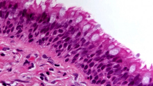
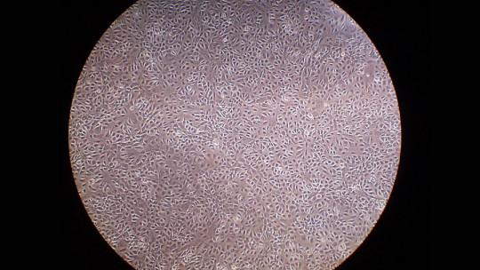
As we said, Keratinocytes are the most abundant cell type in the human epidermis In our species, they make up 95% of the cell bodies in this layer, accompanied in a much smaller proportion by melanocytes, Langerhans cells and Merkel cells.
As its name indicates, keratinocytes They are responsible for synthesizing keratin and, in turn, giving the pertinent properties to each of the four layers of the epidermis: basal layer, stratum spinosum, stratum granulosum and stratum corneum. As a curiosity, it should be noted that the passage of a cell from the basal layer to the horny area lasts about 15 days, an extremely fast period if we look at the rate of tissue turnover in other parts of the body.
Characteristics of keratinocytes
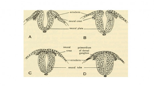
First of all, it should be noted that This cell type has ectodermal origin, that is, it comes from the outermost distal layer of the fetus and the first to develop They are a cell type that releases very little matrix and, therefore, the membranes of adjacent cells are tightly bound. This makes all the evolutionary sense in the world: the less space left between the bricks of a wall, the more difficult it will be for cracks to appear.
In addition to physical proximity, it should be noted that between keratinocytes there are a series of junctions called desmosomes. This “bridge” is mediated by cadherins to a series of intermediate filaments (keratin), which allow the union between cells, thus giving the epidermis a cohesion and integrity that is very resistant over time.
The classic keratinocyte is made up of 72-80% water, cytoplasm, organelles, nucleus and the expression of various types of keratin depending on your location.
It is necessary to note, at this point, that keratinocytes do not have a specific shape throughout their entire life (which in humans is one month), since they go through different epidermal layers and, therefore, they must adapt to different functionalities. In order to show you what these cell bodies are like in each stage and stratum, we must show you, even in general terms, what the keratinization process consists of. Go for it.
Keratinization in a nutshell
The terminal differentiation of keratinocytes from the stratum basale to the corneum occurs under a process known as “keratinization.” We will see its particularities in a superficial way layer by layer.
1. Basal layer
It is the first layer of the epidermis, specifically, the only one in which melanocytes occur, more or less at a rate of one for every 23 keratinocytes. This stratum is conceived as a true tissue factory, since it is formed by only a row of keratinocytes that divide with the purpose of populating the following layers.
These keratinocytes are attached to the basal lamina (which separates the dermis from the epidermis) through hemidesmosome-type junctions, so one cell pole is clearly differentiated from the other. We do not want to go into histological particularities, but it is enough to know that adult epithelial stem cells are found in this stratum, which give rise to keratinocytes. To give you an idea, there is usually one stem cell for every 3,500 keratinocytes in this layer.
2. Stratum spinosum
It originates from the mitotic division of cells in the stratum basale, so it is found immediately above it In this section, the keratinocytes adopt a polyhedral shape about 15 micrometers in diameter, larger and more turgid than those present in the stratum basale. The name “spiny” comes from the desmosome-type junctions and tonofibrils that connect the cells to each other.
It should be noted that, as they advance through the strata, keratinocytes express different cytoplasmic proteins of the keratin type. If K5 and K14 dominated in the basal stratum, here we find K1 and K10.
3. Stratum granulosum
In this layer an important event takes place: gene expression (the synthesis of substances encoded by nuclear DNA) of keratinocytes changes. In the stratum granulosum, these cell types synthesize keratohyalin granules, basophilic compounds of an irregular nature that occur in the cytoplasm of the keratinocytes in this layer (hence their name). The typical keratin types of this phase are K2 and K11.
4. Stratum corneum
In the stratum corneum, keratinocytes differentiate and degenerate, giving rise to corneocytes These do not have a nucleus or cytoplasmic organelles: they only have a thick membrane and multiple lipids, necessary for the structure. They are approximately 50 micrometers in diameter (they are larger than the rest) and are organized in columns of 10 to 30 units to form the stratum corneum itself.
It should be noted that, in addition to losing the nucleus and organelles, corneocytes do not retain more than 15% water by weight, compared to 70% for keratinocytes in the basement membrane. This gives the outermost layer of the epidermis its necessary dryness, which is very important for many microorganisms to be unable to colonize it.
Its relationship with the immune system
As you can see, One of the most striking functions of keratinocytes is to “die” to become insurmountable biological barriers but this is not the only one of its essential tasks.
It should be noted that the invasion of pathogens or contact with allergens in the epidermis brings out the more “immune” side of the keratinocytes. These produce a plethora of cytokines, proteins of a pro-inflammatory nature, such as interleukins (IL)-1, -6, -7, -8, -10, -12, -15, -18, and -20. These cytokines attract immune bodies such as monocytes or T lymphocytes to the site, which begin to act and/or divide in order to destroy the pathogenic agent.
Pathologies known to everyone, such as contact dermatitis, rest on this physiological basis When the immune system recognizes a harmless allergen as harmful, lymphocytes travel to the surface of the skin and produce local responses, such as the characteristic itching, swelling and rash. Although it may not seem like it, the immune system is fighting an unfounded pathogen.
Summary
As you have seen, keratinocytes have both a direct and indirect protective function. Not only are they structural “plugs” in their final phase due to their large size and low water content, but they are also capable of secreting substances that warn the immune system that something is wrong and promote the appearance of local reactions, for the better. and for worse.
Can you imagine what would have happened to human beings without a series of mechanisms as effective as those presented here? In a three-dimensional environment in which even the sun attacks us, understanding life without keratinocytes and the epidermis would be an impossible task.

