


See more of our articles in your search results.
You stare at a photograph of your childhood home and, for a split second, nothing registers — just shapes and colors that refuse to resolve into memory. Or maybe you’ve had a migraine that left you seeing shimmering zigzags in the corner of your vision, and you wondered, with a flicker of fear, whether something serious was happening inside your skull. Vision feels automatic until the moment it isn’t, and that moment can be genuinely unsettling. Understanding the small structure behind these experiences — the occipital lobe — can turn that fear into informed curiosity.
It matters because vision is not a passive act of “seeing what’s there.” It is an active, layered process of construction, and when something interrupts it, the effects ripple into memory, mood, safety, and identity. People who experience visual field defects, sudden visual hallucinations, or difficulty recognizing familiar faces often feel isolated, confused, or embarrassed, unsure whether to mention it to a doctor or dismiss it as stress. Caregivers of stroke survivors frequently describe watching a loved one bump into doorframes on one side, unaware anything has changed, which is disorienting for everyone involved. Knowing that these experiences map onto a specific, well-studied region of the brain can replace shame with understanding.
So what exactly is happening back there, at the rear of your skull, every time you glance at a sunset or read this sentence?
This article walks through the anatomy, structure, and everyday functions of the occipital lobe, along with what happens when this visual processing center is disrupted by injury, disease, or neurological conditions.
What Is the Occipital Lobe and Where Is It Located?
The occipital lobe is the smallest of the brain’s four paired lobes, sitting at the very back of the skull beneath the occipital bone. It is the brain’s dedicated visual processing center, responsible for turning raw light signals into the coherent, three-dimensional world you experience every waking moment. Despite occupying only about 12 to 18 percent of the cerebral cortex’s total surface area, its influence over daily functioning is enormous.
How many of these have you been "meaning to read" for months?
- 1
 TarcherThe Artist's Way30th Anniversary EditionListen Free On Audible
 - 2
 Atria BooksThe Courage to Be DislikedThe Japanese Phenomenon That Shows You How to Change Your Life and Achieve Real HappinessListen Free On Audible
 - 3
 Hay HouseBecoming SupernaturalHow Common People Are Doing the UncommonListen Free On Audible
Anatomically, the occipital lobe sits posterior to the parietal and temporal lobes, separated from them by the parieto-occipital sulcus, and it rests just above a sheet of tissue called the tentorium cerebelli, which divides it from the cerebellum below. Like every other lobe, it exists as a mirrored pair — one on the left hemisphere, one on the right — connected across the midline. Each side processes visual information from the opposite half of the visual field, a crossover pattern that becomes critically important when clinicians interpret visual field defects after a stroke or brain injury.
Blood supply to this region comes primarily from the posterior cerebral artery, a detail that matters clinically because strokes affecting this specific vessel produce a recognizable pattern of vision loss rather than the weakness or speech changes associated with strokes elsewhere. Physiotherapy and neurology resources describe the occipital lobe’s boundaries as somewhat arbitrarily defined compared to other lobes, since there is no single dramatic groove separating it from its neighbors the way the central sulcus separates the frontal and parietal lobes.
Even though it is compact, the occipital lobe does not work in isolation. It constantly exchanges information with the parietal lobe for spatial orientation and with the temporal lobe for object identification and memory, making it one of the most densely interconnected regions in the entire brain.
How Is the Occipital Lobe Structured Internally?
Internally, the occipital lobe is organized into distinct gyri, sulci, and cytoarchitectural zones known as Brodmann areas, each handling a slightly different stage of visual analysis. This layered structure is why damage to one small area can produce a very specific symptom rather than total blindness.
The most important structural landmark is the calcarine sulcus, a deep groove running along the medial surface of the lobe. The cortex lining this sulcus forms the primary visual cortex, also called Brodmann area 17 or V1. Surrounding this core region are the visual association areas, Brodmann areas 18 and 19, which correspond to what researchers now label V2, V3, and V4. On the lateral surface, the superior, middle, and inferior occipital gyri contribute additional processing capacity, while the cuneus and lingual gyrus occupy the medial surface above and below the calcarine sulcus respectively.
- The calcarine sulcus houses the primary visual cortex and is the first cortical stop for signals arriving from the retina.
- The cuneate gyrus sits above the calcarine sulcus and contributes to processing the lower half of the visual field.
- The lingual gyrus lies below it and helps process the upper visual field along with some color-related functions.
- The visual association cortex (Brodmann areas 18 and 19) integrates raw visual data into recognizable patterns, objects, and scenes.
This tiered architecture — raw signal in the calcarine sulcus, meaning-making in the surrounding association cortex — mirrors how neuroscientists Nobel laureates David Hubel and Torsten Wiesel first described visual processing in their landmark work on cortical neurons, showing that individual cells in this region respond selectively to lines, edges, and orientations rather than to whole images at once.

What Does the Primary Visual Cortex Actually Do?
The primary visual cortex, or V1, is the brain’s first stop for interpreting light, converting raw retinal signals into basic building blocks like edges, orientation, and contrast before passing them along for higher-level interpretation. Without a functioning V1, conscious visual awareness essentially disappears, even if the eyes themselves are perfectly healthy.
Light enters the eye, strikes the retina, and travels along the optic nerve, through the optic chiasm and thalamus, before arriving at V1 as electrical impulses rather than anything resembling a picture. V1 neurons are exquisitely tuned to detect specific features: some fire only in response to horizontal lines, others to vertical lines, others to particular angles of movement. This granular, feature-by-feature decoding is what Hubel and Wiesel’s experiments on the visual cortex famously revealed, earning them the Nobel Prize and reshaping how scientists understand sensory processing generally.
From V1, information fans out into the surrounding visual association cortex, where increasingly complex features get assembled — first simple shapes, then textures, then whole objects and faces. This is why a lesion confined strictly to V1 tends to produce a blind spot in a specific part of the visual field (a scotoma) rather than a total inability to recognize objects, while damage further downstream produces more selective deficits like visual agnosia, the inability to recognize objects despite intact basic vision.
One practical takeaway: because V1 damage produces such precisely mapped visual field losses, neurologists can often localize a brain injury or stroke with remarkable accuracy just by charting exactly which part of a patient’s vision has gone dark.
What Are the Dorsal and Ventral Visual Streams?
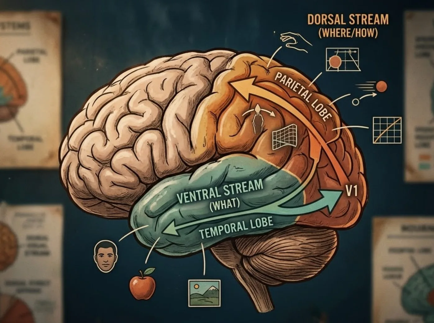
Vision splits into two parallel processing pathways once it leaves the occipital lobe: a “where” pathway for spatial location and movement, and a “what” pathway for object identity. This division, known as the dual visual stream theory, explains why some brain-damaged patients can reach accurately for an object they insist they cannot consciously see.
The dorsal stream travels upward from the occipital lobe into the parietal lobe and handles spatial awareness, motion detection, and the visual guidance of movement — essentially answering “where is it and how do I interact with it.” The ventral stream travels downward into the temporal lobe and specializes in object and face recognition, answering “what am I looking at.” This influential framework was developed by researchers Melvyn Goodale and David Milner, whose case studies of patients with selective brain damage demonstrated that these two systems can be dissociated from one another.
A famous illustration comes from patients with a condition affecting the ventral stream who can accurately post a letter through a mail slot at any angle (dorsal stream intact) while being completely unable to consciously describe the slot’s orientation (ventral stream impaired). This dissociation underscores a broader truth: visual perception and visual action are not the same cognitive process, even though they feel seamless in everyday life.
For readers navigating a loved one’s recovery from occipital or parietal injury, understanding these two streams can help make sense of confusing symptoms, like a person who can dodge an obstacle while walking but cannot verbally identify what the obstacle was.
How Does the Brain Perceive Color, Depth, and Motion?
Color, depth, and motion are each handled by specialized clusters of neurons within the visual association cortex, not by the eyes themselves. The eyes merely detect wavelengths of light and differences in retinal disparity; the occipital lobe does the interpretive work that turns those raw signals into a rich, layered visual experience.
Color perception relies heavily on an area sometimes referred to as V4, which processes signals from cone cells in the retina and compares wavelength information across the visual field to maintain color constancy — the reason a red apple still looks red under both fluorescent light and sunlight. Neuroscientist Semir Zeki’s research on the visual brain was instrumental in mapping how specialized these color-processing regions are, showing that damage restricted to this area can produce a rare condition called achromatopsia, a total loss of color vision despite otherwise normal sight.
Depth and distance perception depend on the brain comparing slightly different images from each eye (binocular disparity) alongside monocular cues like relative size and motion parallax. Motion detection, meanwhile, is concentrated in an area often labeled V5 or MT, located near the boundary of the occipital and temporal lobes; damage here can produce akinetopsia, a bizarre condition in which the world appears as a series of disconnected still frames rather than smooth motion.
- Color processing depends on specialized neurons that compare wavelength across the visual field for consistent hue perception.
- Depth perception combines input from both eyes with cues like size and parallax to build a three-dimensional map.
- Motion detection relies on dedicated cells that track speed and direction, distinct from the circuits handling static shape.
How Does the Occipital Lobe Connect Vision to Memory and Language?
Vision rarely stands alone; the occipital lobe constantly hands information to memory and language networks so that what you see becomes meaningful rather than just decorative. This integration is why recognizing a friend’s face instantly triggers their name, your last conversation, and an emotional response, all within a fraction of a second.
The ventral visual stream connects directly into the medial temporal lobe, an area central to memory formation and long studied by memory researcher Brenda Milner, whose work with patients who had temporal lobe damage helped establish how visual recognition and episodic memory are linked but separable systems. When this visual-to-memory pathway is disrupted, people can retain the ability to see fine detail while losing the ability to attach meaning or familiarity to what they observe — a distressing experience sometimes described as looking at a stranger’s face where a loved one’s should be.
Reading also depends on this occipito-temporal connection. A region near the border of these two lobes, sometimes called the visual word form area, specializes in recognizing letter strings as words rather than arbitrary shapes. Damage nearby can produce alexia, a selective loss of reading ability in someone who can still write and speak fluently — a striking demonstration of how narrowly specialized visual processing circuits can be.
For anyone supporting a family member recovering from a stroke, this explains why speech therapy after occipital or occipito-temporal damage often needs to specifically target reading and object-naming exercises rather than assuming general language recovery will restore these skills automatically.

What Happens When the Occipital Lobe Is Damaged?
Damage to the occipital lobe most commonly produces some form of vision loss, ranging from a small blind spot to complete cortical blindness, depending on how much tissue is affected and on which side. Common causes include stroke, traumatic brain injury, tumors, and, less commonly, degenerative disease affecting posterior brain regions.
Because each occipital lobe processes the opposite half of the visual field, damage confined to one side typically produces homonymous hemianopia — a loss of the same side of vision in both eyes rather than blindness in just one eye. This distinction often surprises patients, who assume vision loss must correspond to a single damaged eye rather than a damaged brain region. Bilateral occipital damage, though rarer, can cause cortical blindness, a total loss of visual awareness despite structurally healthy eyes.
One particularly striking and under-recognized condition is Anton’s syndrome, in which a person with cortical blindness confidently insists they can still see, confabulating explanations for their frequent collisions with furniture. Neurologist Oliver Sacks documented numerous cases of unusual visual disturbances following brain injury in his writing, helping bring public understanding to just how strange and specific occipital lobe damage can look compared to the popular imagination of “blindness.”
- Homonymous hemianopia causes loss of the same visual field on both eyes after one-sided occipital damage.
- Cortical blindness results from extensive bilateral damage despite otherwise healthy eyes.
- Visual agnosia leaves basic sight intact but disrupts the ability to recognize objects or faces.
- Anton’s syndrome involves a striking denial of blindness alongside confident, false descriptions of the environment.
What Conditions and Disorders Involve the Occipital Lobe?
Several distinct neurological and psychiatric conditions trace back to disrupted occipital lobe function, ranging from migraine to rare psychiatric syndromes involving perception. Recognizing these patterns helps separate ordinary eye strain from something that warrants a medical evaluation.
Occipital lobe epilepsy produces seizures that begin with visual symptoms — flashing lights, colored spots, or formed hallucinations — before potentially spreading to other brain regions and producing more typical convulsive symptoms. Migraine with aura similarly originates from waves of altered electrical activity spreading across the occipital cortex, producing shimmering zigzag lines or blind spots that typically resolve within an hour, distinguishing them from the more sustained deficits seen after stroke.
Prosopagnosia, or face blindness, is another occipito-temporal condition in which a person cannot recognize familiar faces, sometimes including their own reflection, despite normal intelligence and otherwise intact vision. Some cases are acquired through brain injury, while others appear to be developmental, present from an early age without any identifiable damage. Psychiatrist and neurologist Antonio Damasio’s research on visual recognition deficits was foundational in distinguishing prosopagnosia from more general memory or intellectual impairment, clarifying that it reflects a specific breakdown in facial processing circuitry rather than a broader cognitive decline.
Charles Bonnet syndrome deserves special mention because it frequently frightens older adults with significant vision loss: as the eyes send less information to the occipital lobe, the brain sometimes fills the gap with vivid, complex visual hallucinations of people, animals, or patterns. Importantly, people experiencing this retain full insight that the images are not real, distinguishing it clearly from a psychotic disorder, though the experience itself can be alarming without proper explanation from a healthcare provider.

How Are Occipital Lobe Conditions Diagnosed and Managed?
Diagnosis typically begins with a detailed description of the visual symptoms, since the pattern and timing of vision changes often points clinicians directly toward the occipital lobe before any imaging is even done. A neurologist will usually map the visual field carefully, since the shape of a blind spot is diagnostically meaningful in a way that “blurry vision” alone is not.
Imaging tools such as MRI and CT scans confirm structural causes like stroke, tumor, or hemorrhage, while electroencephalography (EEG) helps identify occipital lobe epilepsy by capturing abnormal electrical discharges during or between seizures. Functional MRI studies, building on the foundational work of vision researchers like Semir Zeki and the two-streams framework established by Goodale and Milner, have also become valuable research tools for mapping exactly which sub-regions are involved in a given patient’s deficit.
Management depends entirely on the underlying cause. Stroke-related vision loss often benefits from structured vision rehabilitation therapy, which trains patients to compensate through eye-scanning techniques and environmental adaptations rather than expecting full spontaneous recovery. Occipital lobe epilepsy is generally managed with anti-seizure medication, and migraine with aura is addressed through both preventive and acute treatment strategies tailored to trigger patterns.
- Seek prompt evaluation for any sudden vision change, since early treatment for stroke-related causes significantly affects long-term outcomes.
- Track symptom patterns such as timing, duration, and accompanying headache, which helps clinicians distinguish migraine aura from more serious causes.
- Engage in vision rehabilitation exercises consistently, since compensatory scanning strategies improve functional independence over time.
- Communicate hallucinatory experiences openly with a provider, since conditions like Charles Bonnet syndrome are benign but often go unreported out of fear or embarrassment.
How Does the Occipital Lobe Change Across the Lifespan?
Visual processing capacity is not fixed; the occipital lobe develops rapidly in infancy and can show measurable decline or resilience in older adulthood depending on overall brain health. Understanding this trajectory helps set realistic expectations for both childhood milestones and age-related visual changes.
In infants, the primary visual cortex is still maturing, which is why newborns have limited depth perception and poor visual acuity that sharpens dramatically over the first year of life as neural connections in this region strengthen through experience-dependent plasticity. Children with congenital visual impairments benefit enormously from early intervention precisely because this developmental window offers the greatest capacity for the visual cortex to adapt.
In older adults, normal aging can bring subtle declines in contrast sensitivity and motion detection tied to gradual changes in occipital lobe function, distinct from separate eye-related conditions like cataracts or macular degeneration. Conditions such as posterior cortical atrophy, an atypical variant of dementia that begins with visual symptoms rather than memory loss, specifically target this brain region and can be misdiagnosed initially as an ophthalmologic problem rather than a neurodegenerative one. Recognizing that a persistent, unexplained difficulty with reading, navigating stairs, or judging distances in an older adult could reflect posterior cortical atrophy — rather than simply “getting older” — can prompt a more accurate and timely diagnosis.
Supporting healthy visual processing across the lifespan includes protecting the head from repeated trauma, managing cardiovascular risk factors that affect blood flow to the posterior cerebral artery, and staying visually and cognitively engaged, all of which contribute to the resilience of this remarkable but vulnerable part of the brain.
FAQs about the Occipital Lobe
What is the main function of the occipital lobe?
The occipital lobe’s main function is visual processing — interpreting raw signals from the eyes and transforming them into recognizable shapes, colors, motion, and depth. It houses the primary visual cortex, which handles basic feature detection, along with surrounding association areas that build increasingly complex representations of the visual world. Beyond simple sight, it works closely with the parietal and temporal lobes to support face recognition, reading, spatial navigation, and memory formation. Without a functioning occipital lobe, a person can have perfectly healthy eyes and still experience significant, sometimes total, loss of conscious visual awareness.
What happens if the occipital lobe is damaged?
Damage to the occipital lobe typically causes some degree of vision loss, and the specific pattern depends on exactly where and how much tissue is affected. One-sided damage often produces homonymous hemianopia, a loss of the same half of the visual field in both eyes, while extensive bilateral damage can cause cortical blindness despite structurally normal eyes. Some patients also experience visual agnosia, an inability to recognize familiar objects or faces, or rare conditions like Anton’s syndrome, where a person denies being blind at all. The exact symptoms depend heavily on which specific sub-region, from the primary visual cortex to the surrounding association areas, sustains the injury.
Can occipital lobe damage cause hallucinations?
Yes, occipital lobe dysfunction can trigger visual hallucinations, and these appear in several distinct forms. Occipital lobe seizures often begin with simple hallucinations like flashing lights or colored shapes before potentially spreading further. Charles Bonnet syndrome produces vivid, complex hallucinations in people with significant vision loss, even though they retain full awareness that the images are not real. Migraine with aura can also cause temporary visual disturbances like shimmering zigzag patterns. None of these forms of occipital-related hallucination indicate psychosis, since insight into their unreal nature typically remains intact.
Where exactly is the occipital lobe located in the brain?
The occipital lobe sits at the very back of the brain, positioned posterior to the parietal and temporal lobes and directly beneath the occipital bone of the skull. It rests on top of a membrane called the tentorium cerebelli, which separates it from the cerebellum below. Like the other cerebral lobes, it exists as a mirrored pair, with a left and right occipital lobe divided along the brain’s midline. Each side is responsible for processing visual information from the opposite half of the visual field, a crossover pattern that becomes clinically significant when doctors interpret vision loss after a stroke.
Is occipital lobe epilepsy dangerous?
Occipital lobe epilepsy is generally manageable with appropriate medical care, though it requires proper diagnosis since its visual symptoms can be mistaken for migraine or eye problems. Seizures originating here often start with visual phenomena, such as flashing lights, colored spots, or brief blind spots, sometimes progressing to more typical convulsive seizure activity if the electrical disturbance spreads to other brain regions. Left undiagnosed, recurrent seizures can affect quality of life and, in rare cases, pose safety risks during episodes involving loss of awareness. Anti-seizure medication, guided by a neurologist and often supported by EEG monitoring, is the standard and generally effective approach to management.
Can you recover vision after occipital lobe damage?
Some recovery is possible, particularly in the weeks and months following an acute injury like a stroke, though the degree varies significantly between individuals. Vision rehabilitation therapy, which trains compensatory scanning strategies and makes practical use of the intact visual field, has demonstrated meaningful improvements in daily functioning even when the underlying visual field defect itself does not fully resolve. Younger patients and those with smaller areas of damage generally show greater capacity for improvement, reflecting the brain’s overall neuroplasticity. Working closely with a neurologist and a specialized vision rehabilitation therapist gives patients the best realistic chance at regaining functional independence.
How is the occipital lobe different from the visual cortex?
The terms are related but not identical: the occipital lobe is an anatomical region, while the visual cortex refers to the functional tissue that processes vision, most of which happens to sit inside the occipital lobe. The primary visual cortex, or V1, along with the surrounding visual association areas, make up the bulk of the occipital lobe’s tissue. However, visual processing does not stop there — it extends into parts of the neighboring parietal and temporal lobes as information travels along the dorsal and ventral visual streams. So while nearly everything inside the occipital lobe is devoted to vision, not everything involved in vision is confined strictly to the occipital lobe.
Bibliography
- StatPearls Publishing. (2023). Neuroanatomy, Occipital Lobe. National Center for Biotechnology Information Bookshelf.
- Cleveland Clinic. (2022). Occipital Lobe: Function, Location & Conditions.
- Kenhub. (2021). Occipital Lobe: Anatomy, Function and Clinical Relations.
- Physio-pedia. (2025). Occipital Lobe.
- Hubel, D. H., & Wiesel, T. N. (2004). Brain and Visual Perception: The Story of a 25-Year Collaboration. Oxford University Press.
- Sacks, O. (1985). The Man Who Mistook His Wife for a Hat and Other Clinical Tales. Summit Books.
- Milner, A. D., & Goodale, M. A. (1995). The Visual Brain in Action. Oxford University Press.
- Zeki, S. (1993). A Vision of the Brain. Blackwell Scientific Publications.
- Damasio, A. R. (1990). Category-related recognition defects as a clue to the neural substrates of knowledge. Trends in Neurosciences.
- World Health Organization. Stroke, Cerebrovascular Accident.
Use this citation format to reference the article clearly and help readers find the original source.
PsychologyFor. (2026). Occipital Lobe: Anatomy, Characteristics and Functions. PsychologyFor. https://psychologyfor.com/occipital-lobe-anatomy-characteristics-and-functions/