

See more of our articles in your search results.
I remember the first time I had to explain cortical atrophy to a patient’s family. They’d just received the MRI results for their 68-year-old mother, and the radiologist’s report mentioned “cortical atrophy consistent with age-related changes.” The daughter looked at me with fear in her eyes and asked, “Does this mean my mother has Alzheimer’s?” I had to explain something that’s both simple and complicated: yes, her brain was shrinking, but no, that didn’t necessarily mean dementia was inevitable. Cortical atrophy is one of those neurological findings that sounds terrifying but requires careful context to understand what it actually means for any individual person.
The cerebral cortex is the outer layer of the brain—the wrinkled, folded surface you see in pictures of brains. It’s where most of our higher-level thinking happens: language, memory, reasoning, sensory processing, voluntary movement. When we talk about cortical atrophy, we’re describing the progressive loss of neurons and the connections between them in this critical brain region. The cortex literally shrinks, the grooves between the folds widen, and the brain’s overall volume decreases.
Here’s what makes cortical atrophy both fascinating and frightening from a neurological perspective: some degree of brain atrophy is completely normal with aging. Everyone’s brain shrinks somewhat as they get older. But when atrophy happens too quickly, too extensively, or in specific patterns, it signals something pathological—a disease process actively destroying brain tissue. The challenge is distinguishing between normal aging and disease, between changes that are concerning and those that are expected.
So let’s dive deep into cortical atrophy—what it looks like, what causes it, which diseases are associated with it, and what can be done about it. This isn’t just academic neurology. Understanding cortical atrophy matters because it’s increasingly being detected on brain scans, and people need to understand what these findings mean for their cognitive future and quality of life.
What Exactly Is Cortical Atrophy?
Cortical atrophy refers specifically to the loss of neurons and neural connections in the cerebral cortex, as opposed to other brain regions. The brain can be divided roughly into gray matter (the cell bodies of neurons, concentrated in the cortex) and white matter (the connections between neurons). Cortical atrophy primarily affects gray matter, causing the outer surface of the brain to thin and shrink.
How many of these have you been "meaning to read" for months?
- 1
 Undercover Publishing HouseStop Letting Everything Affect YouHow to break free from overthinking, emotional chaos, and self-sabotage.Listen Free On Audible
 - 2
 Penguin Random HouseAtomic HabitsAn Easy & Proven Way to Build Good Habits & Break Bad OnesListen Free On Audible
 - 3
 Harriman HouseThe Psychology of MoneyTimeless lessons on wealth, greed, and happinessListen Free On Audible
This shrinkage happens through multiple mechanisms. Neurons die, either through normal apoptosis (programmed cell death) or through pathological processes. The dendrites and axons connecting neurons to each other retract and disappear. The synapses where neurons communicate deteriorate. Blood vessels in the cortex may narrow or become damaged. The supporting glial cells that maintain brain health may become dysfunctional. All of these processes result in less brain tissue occupying the skull.
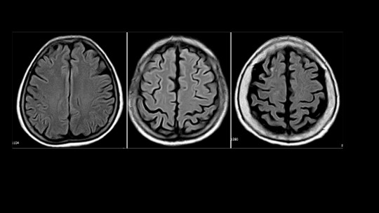
On brain imaging like MRI or CT scans, cortical atrophy shows up in characteristic ways. The sulci—the grooves between the brain’s folds—become wider and more prominent as the brain tissue shrinks away from them. The ventricles—fluid-filled spaces inside the brain—expand to fill the space left by shrinking brain tissue. The cortex itself appears thinner when measured. Radiologists can quantify these changes and compare them to what’s expected for someone’s age.
Cortical atrophy can be classified in several ways. It can be generalized (affecting the whole cortex) or focal (concentrated in specific regions). It can be symmetric (affecting both brain hemispheres equally) or asymmetric (worse on one side). The pattern and location of atrophy often provides clues about what’s causing it and what symptoms might develop.
Symptoms of Cortical Atrophy
The symptoms of cortical atrophy depend entirely on which parts of the cortex are affected and how severely. The cortex isn’t one uniform structure—different regions specialize in different functions. When atrophy damages specific cortical areas, the functions those areas controlled begin to deteriorate.
If atrophy affects the frontal cortex, symptoms might include changes in personality and behavior, difficulty with planning and organization, problems with impulse control, reduced motivation, and changes in social appropriateness. The frontal lobes are our executive control center, so their deterioration affects our ability to regulate ourselves and plan for the future.
When atrophy damages the temporal cortex, memory problems become prominent. The temporal lobes, particularly the hippocampus and surrounding structures, are critical for forming new memories and retrieving old ones. People might repeat questions, forget recent conversations, get lost in familiar places, or struggle to remember names and words. Language comprehension can also suffer since language processing centers reside in the temporal cortex.
Atrophy in the parietal cortex causes problems with spatial awareness, sensory processing, and mathematical reasoning. People might have trouble judging distances, difficulty with coordination and using tools, problems with reading and writing, and confusion about spatial relationships. They might not recognize objects by touch or have trouble integrating information from different senses.
The occipital cortex processes visual information, so atrophy here produces visual symptoms despite the eyes themselves working fine. This is what happens in posterior cortical atrophy—people have trouble reading, recognizing faces or objects, judging distances, distinguishing moving from stationary objects, and navigating visually complex environments. They might see perfectly well but can’t make sense of what they’re seeing.
Generalized cortical atrophy affecting multiple regions produces a broader constellation of symptoms. People might experience cognitive decline across multiple domains—memory, language, reasoning, and judgment all deteriorating together. Physical symptoms like tremors, difficulty walking, stiffness, and coordination problems can emerge if atrophy extends to motor areas. Behavioral and personality changes, mood disturbances, and loss of independence in daily activities often follow as the atrophy progresses.
Causes of Cortical Atrophy
Understanding what causes cortical atrophy requires distinguishing between normal aging and pathological processes. Some degree of brain shrinkage is expected as we age—neurons naturally die, connections are lost, and brain volume decreases gradually. But when atrophy exceeds what’s normal for age, disease processes are usually responsible.
Normal Aging
Even in healthy aging, the brain loses volume at a rate of approximately 0.5% per year after age 60, though rates vary considerably between individuals. This age-related atrophy is generally symmetric, gradual, and doesn’t produce dramatic cognitive impairment. People maintain their independence and cognitive function despite some brain shrinkage. The key is that the atrophy progresses slowly and affects the entire brain relatively evenly.
Neurodegenerative Diseases
The most common cause of pathological cortical atrophy is neurodegenerative disease—conditions where neurons progressively die due to accumulated toxic proteins or other cellular dysfunction. Alzheimer’s disease is the leading culprit, accounting for the majority of cases where cortical atrophy exceeds normal aging. In Alzheimer’s, abnormal proteins called amyloid plaques and tau tangles accumulate in the cortex, triggering inflammation and neuronal death. The atrophy typically begins in the temporal lobes, affecting memory first, then spreads to other regions.
Frontotemporal dementia causes prominent atrophy in the frontal and temporal lobes, producing dramatic personality changes, behavioral disinhibition, and language problems often before significant memory loss. Lewy body dementia, caused by alpha-synuclein protein accumulation, produces cortical atrophy along with visual hallucinations, fluctuating cognition, and movement problems similar to Parkinson’s disease.
Huntington’s disease, a genetic disorder, causes atrophy that begins in deep brain structures but eventually extends to the cortex. Corticobasal degeneration specifically targets the cortex and basal ganglia, producing asymmetric symptoms with stiffness, poor coordination, and cognitive changes. Multiple system atrophy affects multiple brain regions including cortical areas.
Vascular Causes
Vascular disease—problems with blood vessels supplying the brain—can cause cortical atrophy. Multiple small strokes, chronic reduced blood flow, high blood pressure, and diabetes all damage brain tissue over time. The cortex is particularly vulnerable to vascular insufficiency because it has high metabolic demands. Vascular risk factors like hypertension, high cholesterol, diabetes, and smoking accelerate cortical atrophy even in people without dementia.
Traumatic Brain Injury
Repeated head trauma, as seen in contact sports or military combat, can trigger progressive cortical atrophy years after the injuries. Chronic traumatic encephalopathy (CTE) is characterized by widespread cortical and subcortical atrophy along with accumulation of abnormal tau protein. Even single severe traumatic brain injuries can accelerate atrophy and increase dementia risk.
Infections and Inflammation
Certain infections affecting the central nervous system can cause cortical atrophy. HIV-associated dementia produces generalized brain atrophy including cortical regions. Prion diseases like Creutzfeldt-Jakob disease cause rapid, severe cortical atrophy. Chronic inflammation from autoimmune conditions or persistent infections can gradually damage cortical tissue.
Nutritional and Metabolic Causes
Severe nutritional deficiencies, particularly B vitamins (B1, B12), can contribute to cortical atrophy. Chronic alcoholism causes brain atrophy through multiple mechanisms including nutritional deficiency, direct toxicity, and repeated withdrawal episodes. Anorexia nervosa and other eating disorders can produce significant brain volume loss that may partially reverse with nutritional rehabilitation.
Metabolic conditions like poorly controlled diabetes accelerate cortical atrophy. Thyroid disorders, liver disease, and kidney disease can all affect brain health and contribute to atrophy over time.
Other Causes
Multiple sclerosis, though primarily affecting white matter, can cause cortical atrophy as inflammation damages the cortex. Epilepsy, particularly if poorly controlled, may contribute to progressive atrophy. Certain medications, particularly long-term use of some psychiatric drugs and chronic steroid use, have been associated with brain atrophy. Radiation therapy to the brain can cause delayed atrophy years after treatment.

Disorders Associated with Cortical Atrophy
Let’s look more closely at the specific disorders where cortical atrophy is a prominent feature, because understanding these diseases helps clarify what cortical atrophy means clinically.
Alzheimer’s Disease
Alzheimer’s disease is far and away the most common cause of significant cortical atrophy. The disease typically begins with atrophy in the medial temporal lobes, particularly the hippocampus and entorhinal cortex, which explains why memory problems are usually the first symptom. As the disease progresses, atrophy spreads to other temporal regions, then to parietal and frontal cortex, eventually affecting most of the cortex.
The rate of atrophy in Alzheimer’s is much faster than normal aging—typically 2-3% brain volume loss per year in affected regions compared to 0.5% in healthy aging. This accelerated shrinkage correlates with cognitive decline. On brain imaging, you see prominent atrophy of the hippocampus early on, followed by widening of the sulci and enlargement of the ventricles as the disease progresses.
Posterior Cortical Atrophy
Posterior cortical atrophy is a fascinating variant where atrophy predominantly affects the back of the brain—the posterior parietal and occipital cortex. Most cases are actually caused by Alzheimer’s disease pathology, but it presents very differently because of where the atrophy is concentrated.
People with posterior cortical atrophy develop progressive visual and spatial problems despite having healthy eyes. They struggle with reading, recognizing objects and faces, judging distances, and navigating space. They might have trouble reaching for objects, getting dressed, or telling if things are moving or stationary. Memory often remains relatively preserved early in the disease, unlike typical Alzheimer’s.
This condition typically affects people in their 50s and 60s, somewhat younger than typical Alzheimer’s onset. The visual and spatial symptoms can be misattributed to eye problems initially, delaying diagnosis. On imaging, the atrophy is concentrated posteriorly with relative sparing of frontal and anterior temporal regions early on.
Frontotemporal Dementia
Frontotemporal dementia refers to a group of disorders characterized by prominent atrophy of the frontal and/or temporal lobes. There are several variants. Behavioral variant frontotemporal dementia causes dramatic personality changes, disinhibition, apathy, compulsive behaviors, and loss of empathy due to frontal lobe atrophy. People might become socially inappropriate, develop rigid routines, or lose motivation entirely.
Primary progressive aphasia variants affect language due to temporal lobe atrophy. People gradually lose the ability to speak, understand language, or name objects despite preserved memory and reasoning. The atrophy pattern distinguishes different aphasia types—some affect fluency, others comprehension or word-finding.
Frontotemporal dementia tends to affect people younger than Alzheimer’s—often in the 50s or early 60s. The behavioral changes can be mistaken for psychiatric disorders initially. The prominent frontal and anterior temporal atrophy on imaging helps distinguish it from Alzheimer’s disease.
Corticobasal Degeneration
Corticobasal degeneration causes asymmetric atrophy of the cortex and deeper brain structures called the basal ganglia. It typically begins affecting one side of the body much more than the other. Symptoms include stiffness, slow clumsy movements, tremor, muscle jerks, difficulty with balance, and a characteristic phenomenon where one limb seems to have “a mind of its own” (alien limb syndrome).
Cognitive symptoms include problems with thinking, language, and speech. The progression is gradual but relentless, with increasing disability over years. The disease usually begins between ages 50 and 70. On imaging, you see asymmetric atrophy of the cortex, particularly in frontal and parietal regions, along with atrophy of the basal ganglia on the more affected side.
Lewy Body Dementia
Dementia with Lewy bodies causes cortical atrophy, though often less severe than in Alzheimer’s disease. The distinctive features are fluctuating cognition, visual hallucinations, movement problems like Parkinson’s disease, and sensitivity to antipsychotic medications. The cortical atrophy is often more prominent in posterior regions.
Huntington’s Disease
Huntington’s disease is a genetic disorder causing progressive brain atrophy. While it primarily affects deep brain structures called the basal ganglia and caudate nucleus, cortical atrophy also occurs as the disease progresses. People develop involuntary dance-like movements (chorea), cognitive decline, psychiatric symptoms, and personality changes. The disease typically begins in the 30s or 40s and progresses over 15-20 years.
HIV-Associated Dementia
Before effective antiretroviral therapy, HIV commonly caused progressive dementia with widespread brain atrophy including cortical regions. While much less common now with treatment, HIV can still cause accelerated cognitive decline and atrophy, particularly if infection isn’t well-controlled or treatment is started late.
Multiple Sclerosis
Multiple sclerosis primarily affects white matter through inflammatory demyelination, but cortical atrophy also occurs and correlates with cognitive impairment and disability progression. The cortical damage in MS involves both inflammation and neurodegeneration. Brain atrophy rate is an important marker of MS disease activity and treatment response.
Diagnosis of Cortical Atrophy
Cortical atrophy itself is typically detected through brain imaging—MRI or CT scans. MRI provides much more detailed information and can detect subtle atrophy patterns that distinguish different diseases. Advanced MRI techniques can measure brain volumes precisely and compare them to age-matched norms.
But identifying atrophy on imaging is just one piece of diagnosis. The critical questions are: What’s causing the atrophy? How rapidly is it progressing? What are the functional consequences? And what, if anything, can be done about it?
A comprehensive evaluation includes detailed cognitive testing to characterize what mental functions are affected and how severely. Neuropsychological testing provides objective measures of memory, language, attention, reasoning, and other cognitive domains. This helps distinguish normal aging from pathological decline and tracks progression over time.
Blood tests screen for reversible causes of cognitive impairment and atrophy like thyroid disease, B12 deficiency, and infections. Genetic testing may be appropriate when hereditary conditions like Huntington’s disease or familial Alzheimer’s are suspected. In some cases, cerebrospinal fluid analysis through lumbar puncture can detect biomarkers of Alzheimer’s disease or other conditions.
PET scanning can detect amyloid plaques and tau tangles characteristic of Alzheimer’s disease, helping confirm diagnosis even before severe atrophy develops. FDG-PET shows brain metabolism patterns that differ between various neurodegenerative diseases.
Can Cortical Atrophy Be Treated or Reversed?
This is the question everyone wants answered, and unfortunately, the news is mixed. Currently, there are no treatments that can reverse established cortical atrophy or reliably stop its progression in most neurodegenerative diseases. Once neurons are dead and synapses are lost, we can’t bring them back with any available intervention.
However, that doesn’t mean nothing can be done. Several approaches can help manage symptoms, slow progression in some cases, and maintain quality of life:
Treating Underlying Causes
When cortical atrophy has a treatable underlying cause, addressing it is crucial. Controlling blood pressure, cholesterol, and diabetes can slow vascular contributions to atrophy. Treating vitamin deficiencies, thyroid disorders, or infections can prevent further damage. Stopping alcohol use may allow some partial recovery of brain volume. Managing autoimmune conditions reduces inflammatory damage.
Medications for Specific Diseases
For Alzheimer’s disease, cholinesterase inhibitors (donepezil, rivastigmine, galantamine) and memantine can provide modest symptomatic benefit by enhancing neurotransmitter function, though they don’t stop atrophy. Newer amyloid-targeting medications like lecanemab and donanemab show promise in slowing cognitive decline in early Alzheimer’s by reducing amyloid plaques, though their effects on atrophy are still being studied.
For other conditions, symptomatic treatments address specific problems. Antidepressants help with mood symptoms. Antipsychotics may be used cautiously for behavioral problems. Medications for movement disorders help with tremor and stiffness. The goal is managing symptoms to maintain function and quality of life even as the underlying disease progresses.
Lifestyle Interventions
Multiple lifestyle factors influence brain health and may slow atrophy progression. Regular physical exercise is one of the most robust interventions—it improves cardiovascular health, promotes neuroplasticity, and may reduce atrophy rate. Cognitive stimulation through mentally engaging activities helps maintain cognitive reserve. Social engagement provides cognitive stimulation and emotional support. Good sleep quality is essential for brain health and clearing toxic proteins.
Diet matters too. Mediterranean-style diets rich in vegetables, fruits, whole grains, fish, and healthy fats are associated with better cognitive outcomes and possibly slower atrophy. Managing vascular risk factors through lifestyle and medication is crucial for preventing additional damage.
Cognitive Rehabilitation and Support
Cognitive rehabilitation helps people develop strategies to compensate for declining abilities. Occupational therapy optimizes daily function. Speech therapy addresses language and swallowing problems. Physical therapy maintains mobility and prevents falls. These interventions don’t reverse atrophy but help people function better despite brain changes.
Environmental modifications, assistive devices, and caregiver support become increasingly important as atrophy progresses. The goal shifts from cure to maximizing quality of life, maintaining dignity, and supporting both patients and families through a difficult journey.
Prognosis and What to Expect
The prognosis for cortical atrophy depends entirely on the underlying cause and how rapidly it’s progressing. Age-related atrophy in healthy aging is gradual and compatible with many years of good cognitive function and independence. Some people maintain excellent cognitive health into their 90s despite measurable brain atrophy.
Pathological atrophy from neurodegenerative disease follows a more concerning course. In Alzheimer’s disease, the average time from diagnosis to death is 8-10 years, though this varies widely. Frontotemporal dementia often progresses more rapidly, with average survival of 6-8 years from symptom onset. Corticobasal degeneration typically progresses over 5-10 years with increasing disability.
However, these are averages, and individual trajectories vary considerably. Some people progress rapidly while others remain relatively stable for years. Younger age at onset sometimes means slower progression. Better cardiovascular health, higher education levels, and strong social engagement are associated with better outcomes.
The psychological impact of cortical atrophy diagnosis shouldn’t be underestimated. Learning that your brain is shrinking and your cognitive abilities may decline is devastating. Many people experience anticipatory grief—mourning the future they expected to have. Depression and anxiety are common and need to be addressed as part of comprehensive care.
Research and Future Directions
Neuroscience research into cortical atrophy and its causes is advancing rapidly. Better understanding of the mechanisms driving neurodegeneration is leading to novel therapeutic targets. Biomarkers that can detect disease early, before significant atrophy occurs, may enable intervention when treatments could be most effective.
Advances in neuroimaging allow increasingly precise quantification of atrophy patterns and rates. This helps with diagnosis, prognosis, and monitoring treatment effects in clinical trials. Machine learning algorithms can detect subtle atrophy patterns that distinguish different diseases earlier than human radiologists.
Gene therapy, stem cell treatments, and approaches to clear toxic proteins or enhance neuroplasticity are in various stages of research. While none are proven effective yet, the pace of discovery is accelerating. There’s genuine hope that treatments to slow or prevent cortical atrophy may become available within the next decade.
Living with Cortical Atrophy
For people diagnosed with significant cortical atrophy, the challenge is living fully in the present while planning realistically for the future. This means making legal and financial preparations while still able—advance directives, power of attorney, discussions with family about wishes for care. It means staying engaged with meaningful activities and relationships as long as possible. It means accepting help when needed without giving up autonomy prematurely.
For families and caregivers, it means educating themselves about what to expect, finding support, and taking care of their own health. Caregiving for someone with progressive cognitive decline is emotionally and physically demanding. Respite care, support groups, and professional guidance aren’t luxuries—they’re necessities for sustaining care over months and years.
The key message is this: cortical atrophy is serious and often progressive, but it’s not immediately terminal, and quality of life can be maintained for significant periods with appropriate support and intervention. Understanding what’s happening in the brain helps people make informed decisions and access available resources. While we can’t yet reverse neurodegeneration, we can approach it with knowledge, compassion, and realistic hope.
FAQs About Cortical Atrophy
Is cortical atrophy the same as dementia?
No, cortical atrophy and dementia are not the same thing, though they’re related. Cortical atrophy is a structural finding on brain imaging—physical shrinkage of the cortex. Dementia is a clinical syndrome—significant cognitive decline that interferes with daily functioning. Some degree of cortical atrophy is normal with aging and doesn’t cause dementia. However, when atrophy becomes severe or progresses rapidly, it often causes dementia. Think of atrophy as the physical brain change and dementia as the functional consequence. You can have mild atrophy without dementia, especially in normal aging. But moderate to severe atrophy, particularly if progressing rapidly, usually produces cognitive impairment that may meet criteria for dementia. The relationship between degree of atrophy and cognitive impairment varies—some people maintain surprisingly good function despite significant atrophy (cognitive reserve), while others decline substantially with relatively modest atrophy.
Can you have cortical atrophy without symptoms?
Yes, mild cortical atrophy can exist without noticeable symptoms, especially in normal aging. Many people have mild atrophy detected incidentally on brain scans done for other reasons (like after head injury or for headaches) without any cognitive complaints. The concept of “cognitive reserve” explains this—people with higher education, mentally stimulating careers, and engaged lifestyles can tolerate more brain damage before symptoms emerge because they have more efficient neural networks and compensatory mechanisms. However, as atrophy progresses, symptoms eventually appear. Asymptomatic atrophy may represent a preclinical stage where brain changes are occurring but haven’t yet crossed the threshold to produce noticeable impairment. Regular cognitive monitoring is important for people with more than age-appropriate atrophy even if currently asymptomatic, as symptoms may develop over time.
How quickly does cortical atrophy progress?
The rate of cortical atrophy progression varies dramatically depending on the cause. In healthy aging, brain volume decreases about 0.5% per year after age 60—a slow, gradual process compatible with maintained cognitive function. In Alzheimer’s disease, atrophy rates are much faster, typically 2-3% per year in affected regions, with rates potentially higher in more aggressive cases. Frontotemporal dementia may progress even more rapidly. Vascular dementia progression is often stepwise, with periods of stability punctuated by sudden declines after strokes. Some conditions like Creutzfeldt-Jakob disease cause extremely rapid atrophy over months. The rate can also vary within diseases—some people with Alzheimer’s have slow progression over a decade or more while others decline rapidly over just a few years. Factors affecting progression rate include age at onset, genetics, vascular health, cognitive reserve, and possibly lifestyle factors. Serial brain imaging over time can measure individual atrophy rates and help predict future decline.
Can cortical atrophy be prevented?
You can’t completely prevent age-related cortical atrophy, but you can likely slow it and reduce your risk of pathological atrophy. The same factors that promote cardiovascular health also protect brain health: controlling blood pressure, cholesterol, and blood sugar; not smoking; maintaining healthy weight; exercising regularly; and eating a brain-healthy diet like the Mediterranean diet. Regular physical exercise is particularly important—it’s associated with slower atrophy and lower dementia risk. Staying cognitively and socially engaged appears protective. Getting good sleep, managing stress, avoiding excessive alcohol, and treating depression also matter. Protecting your head from traumatic injuries prevents one cause of accelerated atrophy. Addressing hearing loss may be protective. While these measures don’t guarantee you won’t develop pathological atrophy—some causes like genetic Alzheimer’s disease can’t currently be prevented—they likely reduce risk and slow progression. The strongest evidence is for cardiovascular health and exercise. Starting these preventive measures earlier in life is more effective than waiting until cognitive problems emerge.
What is the difference between cortical and cerebral atrophy?
Cerebral atrophy is the broader term referring to loss of brain tissue anywhere in the cerebrum (the main part of the brain). Cortical atrophy is a specific type of cerebral atrophy affecting primarily the cortex (the outer gray matter). Cerebral atrophy can be cortical (affecting gray matter in the outer layers), subcortical (affecting deep brain structures like basal ganglia or white matter), or both. In cortical atrophy, you see shrinkage mainly in the brain’s surface with widening of the sulci (grooves) between folds. In subcortical or central atrophy, deep brain structures shrink and ventricles enlarge, but the cortical surface may be relatively preserved. Many neurodegenerative diseases cause both cortical and subcortical atrophy, but the predominant pattern helps with diagnosis. Alzheimer’s disease is primarily cortical early on. Conditions like Huntington’s disease start subcortically. The clinical symptoms differ too—cortical atrophy tends to produce cognitive symptoms first, while subcortical atrophy often produces movement problems earlier.
Does cortical atrophy show up on all brain scans?
Cortical atrophy can be detected on both CT scans and MRI, but MRI is much more sensitive and provides more detailed information. Mild atrophy might be missed on CT but visible on MRI. Advanced MRI sequences can quantify brain volumes precisely and detect subtle changes over time. Very early or mild atrophy might not be apparent even on MRI, especially if the radiologist isn’t specifically looking for it or doesn’t have prior scans for comparison. The sensitivity for detecting atrophy also depends on the scanner quality, imaging protocols, and radiologist expertise. Newer techniques using volumetric analysis and comparison to age-matched databases can detect atrophy that might be reported as “normal for age” on visual inspection alone. If there’s clinical concern for cognitive decline, an MRI specifically focused on brain structure with volumetric analysis is the best imaging approach. Serial MRIs comparing brain volumes over time are more sensitive than single scans for detecting progressive atrophy.
Is there any way to regrow brain tissue lost to cortical atrophy?
Unfortunately, with current medical technology, we cannot regrow brain tissue or reverse established cortical atrophy in neurodegenerative diseases. Once neurons are dead and synapses lost, they don’t regenerate. The adult brain has limited neuroplasticity and very limited neurogenesis (creation of new neurons), which occurs mainly in specific regions like the hippocampus. While the brain can sometimes reorganize and recruit other areas to partially compensate for lost tissue, it cannot regenerate substantial lost volume. However, there are important caveats: Some causes of atrophy are partially reversible if caught early and treated—like atrophy from severe malnutrition, alcohol abuse, or certain metabolic disorders. Stopping the damaging process may allow some recovery, though usually not complete restoration. Research into stem cell therapy, gene therapy, and promoting neurogenesis is ongoing, but nothing is proven effective yet for regrowing lost brain tissue. The focus currently is on preventing further loss rather than reversing existing atrophy, which is why early detection and intervention are so important.
Should everyone get brain scans to check for cortical atrophy?
No, routine brain imaging for cortical atrophy isn’t recommended for people without symptoms. There are several reasons: First, mild atrophy is common in normal aging and doesn’t predict dementia reliably. Finding atrophy in an asymptomatic person creates anxiety without clear clinical benefit. Second, there are no proven preventive treatments we’d do differently based on finding mild atrophy in asymptomatic people beyond the lifestyle measures everyone should follow anyway. Third, incidental findings on brain scans often lead to additional testing, costs, and worry without improving outcomes. Brain imaging should be done when there are concerning cognitive or neurological symptoms, when monitoring known disease, or when needed for other medical reasons. If you’re experiencing memory problems, personality changes, difficulty with daily tasks, or other cognitive concerns, then brain imaging along with cognitive testing is appropriate to evaluate for atrophy and other causes. But scanning healthy, asymptomatic people routinely isn’t currently recommended by medical guidelines.
Use this citation format to reference the article clearly and help readers find the original source.
PsychologyFor. (2025). Cortical Atrophy: Symptoms, Causes and Associated Disorders. PsychologyFor. https://psychologyfor.com/cortical-atrophy-symptoms-causes-and-associated-disorders/