Betz Cell: Characteristics And Functions Of This Type Of Neuron
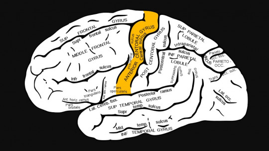
Our brain is responsible for planning, coordinating and executing the movements necessary to carry out daily activities, and it does so mainly through the primary motor area.
In this brain region are some of the largest cells in our nervous system, the Betz cells; a type of giant pyramidal neuron that is responsible for transmitting motor commands through nerve impulses that travel from the neocortex to the spinal cord.
In this article we explain what Betz cells are what are their main characteristics, where they are located, and in what pathological processes they are involved.
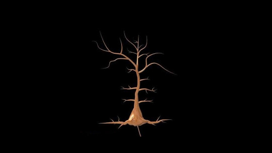
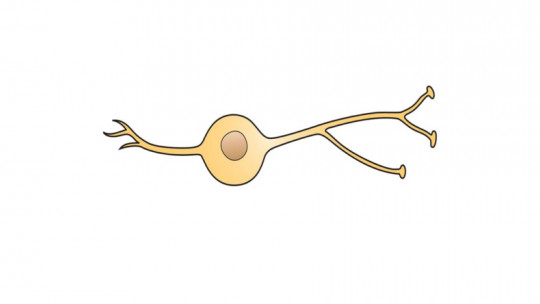
Betz cells are some of the largest motor neurons in the human nervous system , and are named after the Ukrainian scientist Vladimir A. Betz, who described this type of nerve cells at the end of the 19th century. These pyramidal-type cells are giant in size (compared to most neurons) and are located in the gray matter of the primary motor cortex, a brain region responsible, along with other adjacent areas, for planning and executing muscle movements.
Betz neurons are characterized by large somata and extensive basilar dendrites. These dendrites are significantly larger than those of other superficial and deep pyramidal neurons. The apical dendrites and soma of these cells are oriented along a vertical axis, which may contribute to columnar processing in the primary motor cortex. Besides, Betz cell somata have a heterogeneous shape including pyramidal, triangular and spindle-shaped cell bodies.
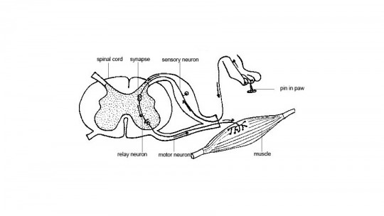
These motor neurons send their axons through the corticospinal tract to the anterior horn of the spinal cord, where they contact the lower motor neuron. Although Betz cells have an apical dendrite typical of pyramidal neurons, they have more primary dendritic axes, and these do not leave the soma only at basal angles, but branch from almost any point in an asymmetric manner.
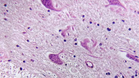
The perisomatic and basal dendrites of Betz neurons project to all cortical layers, but most of its horizontal projections populate layers V and VI , some of which reach the white matter. According to one study, Betz cells represent approximately 10% of the total population of pyramidal cells in layer Vb of the human primary motor cortex.
The primary motor cortex
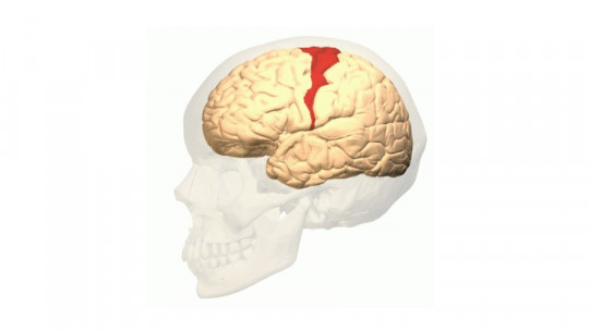
Betz cells are located in layer V of the primary motor cortex. This layer contains this type of giant pyramidal neurons, responsible for sending their long axons to the contralateral motor nuclei of the cranial nerves and to the lower motor neurons located in the ventral horn of the spinal cord.
The axons of Betz neurons are part of the corticospinal tract , and although these nerve cells do not make up the entire motor output of the cortex, they are responsible for providing a clear marker for the primary motor cortex (Brodmann area 4). This region of the brain contains a topographic map of the muscles of our body, in which the head is represented laterally, the leg medially, and the rest of the parts in intermediate positions.
Betz cells are found singly or in small groups of three to four neurons, especially in the dorsal part of the primary motor cortex. The size of the cell bodies of these neurons decreases continuously along a mediolateral gradient. This reduction in size appears to be related to motor somatotopy: the largest cells are found in the region representing feet and legs, where efferent axons project further along the corticospinal tract.
It should be noted that Betz cells They are found in the motor cortex of all primates and, according to studies, the somata of these neurons become proportionally larger with increases in body weight, brain weight, and encephalization. Furthermore, phylogenetic variation in the volumetric scale of this type of neurons could be related to species-specific adaptations.
Neurodegenerative diseases
There appear to be only a few central nervous system pathologies that involve Betz cells. These are, generally, neurodegenerative diseases that more or less specifically affect the primary motor cortex and its projections
The extent to which Betz cells are affected in degenerative motor neuron diseases such as Amyotrophic Lateral Sclerosis (ALS) is still unknown. This progressive disease is known to affect not only the motor system, but also several non-motor systems and subcortical areas, and can occur sporadically or in families. The pathophysiological mechanism in ALS is the loss of anterior horn cells and degeneration of the corticospinal tract with involvement of the upper motor neurons.
There are other neurodegenerative diseases included in the ALS spectrum, for example, the ALS-parkinsonism-dementia complex a disorder involving motor cortical pathways and primary lateral sclerosis involving only upper motor neurons with a complete loss of Betz cells.
At the cellular cortical level, the degeneration of dendritic arborizations, changes in synapses and the loss of Betz cells in ALS and other degenerative diseases that involve the primary motor cortex suggest a participation of this neuronal subpopulation in the process of this type of neurological diseases.
Normal brain aging
Ramón y Cajal was one of the first researchers to identify a difference in Betz cell morphology during lifespan between newborns and adults; the famous anatomist discovered that the basal dendrites of this type of neurons were longer in developed brains
In more recent studies, it has been observed that in normally aging brains, Betz cells have reduced and swollen dendritic spines. These age-related changes have been considered a possible correlate of slowing motor performance and agility, as well as increased stiffness throughout life, as Betz cells They are preferentially involved in the tone of the stabilizing muscles
Furthermore, animal research has reported a decrease in the size of Betz cell somata in normal adult rhesus monkeys, along with a progressive age-related appearance of highly specific inclusion bodies (abnormal subcellular structures). However, these data contradict previous observations of Betz cell inflammation during aging in humans.
The fact that Betz cells can be affected during aging is important considering the fact that studies carried out in this regard have only investigated the brains of elderly patients. All in all, it should be noted that the primary motor cortex is generally spared in Alzheimer’s disease, at least until the very late stages of dementia, and pathological changes in large neurons are only observed in atypical cases with prominent motor symptoms or in cases of the amyotrophic lateral sclerosis-parkinsonism-dementia complex.