


See more of our articles in your search results.
You’re mid-conversation with someone you’ve known for years, and suddenly their name simply isn’t there. Or you catch yourself standing in a room you just walked into, unsure why you came, the memory dissolving before you could grab it. Maybe a loved one had a seizure that left them staring blankly, smacking their lips, and afterward remembering nothing at all — a moment that terrified everyone in the room. These are not signs of failure or weakness; they are windows into one of the most intricate structures in the human brain, the temporal lobe.
It’s easy to feel embarrassed or anxious when memory, language, or emotional regulation seem to misfire, especially when friends or family brush it off as “just getting older” or “not paying attention.” But these functions rely on a densely packed, highly specialized region that manages hearing, language comprehension, emotional processing, and the encoding of new memories, all at once. When that system is disrupted, even slightly, the effects can touch nearly every part of daily life, from holding a job interview to recognizing a spouse’s face across a crowded room. Understanding the biology behind these experiences replaces self-blame with informed compassion.
So what is actually happening inside this part of the brain, and why does it hold so much power over who we are?
This guide explains the anatomy, key regions, functions, and clinical significance of the temporal lobe, including what happens when it is injured, diseased, or affected by seizures.
What Is the Temporal Lobe and Where Is It Located?
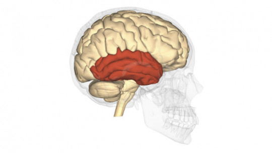
The temporal lobe is one of the four major paired lobes of the cerebral cortex, positioned beneath the lateral or Sylvian fissure, roughly behind the temples and above the ears. It is the second-largest lobe in the human brain, accounting for close to 17 percent of the entire cerebral cortex, and research indicates it reaches its greatest relative size and complexity in humans compared to other primates.
How many of these have you been "meaning to read" for months?


Structurally, the temporal lobe sits inferior to the frontal and parietal lobes and anterior to the occipital lobe, occupying much of what anatomists call the middle cranial fossa at the base of the skull. Like the rest of the cerebral cortex, it exists as a mirrored pair, and in most people the two sides are not functionally identical: the left temporal lobe, dominant in the majority of individuals, specializes in verbal and language-based processing, while the right temporal lobe handles non-verbal information such as music, faces, and spatial-emotional cues.
The temporal lobe divides into three main gyri visible on its lateral surface — the superior, middle, and inferior temporal gyri — each contributing to a different layer of auditory, visual, or semantic processing. Tucked into its medial surface are some of the most clinically important structures in all of neuroscience: the hippocampus, the amygdala, and the surrounding parahippocampal and entorhinal cortices, which together form the medial temporal lobe memory system.
Blood supply arrives chiefly through branches of the middle cerebral artery, with the medial structures additionally served by the posterior cerebral artery — a detail that becomes clinically relevant because strokes or reduced blood flow to this specific territory can selectively impair memory while sparing other cognitive functions.
How Is the Temporal Lobe Organized Into Functional Zones?
The temporal lobe is not a single uniform structure but a mosaic of specialized zones, each handling a distinct cognitive task. Understanding this internal map explains why damage to one small area can produce a very narrow, specific deficit rather than widespread cognitive collapse.
On the superior temporal gyrus sits the primary auditory cortex, corresponding to Brodmann areas 41 and 42, which receives raw sound signals relayed from the ears via the thalamus. Just behind this, in the posterior portion of Brodmann area 22 on the dominant side, lies Wernicke’s area, essential for understanding spoken and written language. The inferior and medial regions, including the fusiform gyrus, specialize in higher-order visual recognition, particularly of faces and complex scenes, working as a bridge between the occipital lobe’s raw visual processing and the temporal lobe’s semantic memory stores.
- The primary auditory cortex decodes pitch, volume, and basic sound patterns arriving from both ears.
- The auditory association area (Brodmann area 22) interprets these raw sounds as meaningful patterns like speech or music.
- The fusiform gyrus specializes in rapid visual recognition, especially of familiar faces.
- The medial temporal lobe, including the hippocampus and amygdala, anchors memory formation and emotional processing.
This zone-based organization was mapped in remarkable detail decades ago by neurosurgeon Wilder Penfield, whose direct electrical stimulation of the temporal lobe in awake epilepsy patients revealed that touching specific points could trigger vivid memory flashbacks, sounds, or even déjà vu sensations, offering some of the earliest direct evidence that memory and perception have identifiable physical locations in this lobe.

How Does the Temporal Lobe Process Sound and Hearing?
Hearing begins in the ears but becomes meaningful sound only once it reaches the temporal lobe. This region transforms vibrations into the words, music, and environmental sounds we consciously perceive and interpret.
Sound waves travel from the cochlea through the auditory nerve and thalamus before arriving at the primary auditory cortex on the superior temporal gyrus, where neurons are organized tonotopically — meaning cells responding to similar frequencies are physically grouped together, much like keys on a piano. From there, the signal moves into surrounding auditory association areas, which layer on interpretation: is this a voice, a car horn, or a melody? The right temporal lobe tends to specialize in interpreting tone, pitch contour, and music, while the left temporal lobe is more heavily weighted toward the rapid, sequential processing required for speech.
Damage restricted to the auditory processing regions can cause auditory agnosia, a rare condition where a person hears sounds perfectly well but cannot identify or interpret what they mean, even though their hearing thresholds test as normal. This distinction matters clinically, because it separates a peripheral hearing problem, which an audiologist would address, from a cortical processing problem, which requires neurological evaluation instead.
A practical takeaway for caregivers: if someone reports hearing sounds clearly but struggling to understand speech specifically in noisy environments or during conversation, this pattern may point toward temporal lobe processing changes rather than simple hearing loss, and it deserves a full neurological work-up rather than just a hearing aid fitting.
What Role Does the Temporal Lobe Play in Language Comprehension?
Language comprehension depends heavily on the temporal lobe, particularly Wernicke’s area, which allows a person to extract meaning from spoken and written words. Without this region functioning properly, speech can sound like a fluent stream of nonsense, both to the listener and, troublingly, to the speaker as well.
Nineteenth-century neurologist Carl Wernicke first identified this region after observing patients who spoke fluently, with normal rhythm and grammar, yet produced sentences devoid of coherent meaning and seemed largely unaware that anything was wrong. This condition, now called receptive aphasia or Wernicke’s aphasia, contrasts sharply with the halting, effortful speech pattern seen in frontal lobe language damage (Broca’s aphasia), illustrating just how specialized different brain regions are even within a single overarching function like language.
The temporal lobe’s involvement in language extends beyond single-word comprehension into the ability to follow complex sentences, understand humor and sarcasm, and process the semantic relationships between concepts. The angular and supramarginal gyri, at the temporal-parietal junction, add another layer, helping integrate auditory and visual language input, which is why damage near this border can selectively impair reading comprehension while sparing spoken language, or vice versa.
For families supporting someone with Wernicke’s aphasia after a stroke, understanding that the person is not being deliberately confusing or careless can meaningfully reduce frustration on both sides, replacing blame with patience and appropriate speech-language therapy referrals.

How Does the Temporal Lobe Store and Retrieve Memory?
Memory formation depends critically on the medial temporal lobe, particularly the hippocampus, which acts as a temporary staging ground that converts everyday experiences into stable, long-term memories. Without it, new experiences can be lived vividly in the moment yet vanish completely within minutes.
The most famous demonstration of this comes from the case of patient H.M., whose hippocampus and surrounding medial temporal structures were surgically removed on both sides to control severe epilepsy. Neuropsychologist Brenda Milner’s decades of research with H.M. revealed that while he could no longer form new conscious, declarative memories, he could still learn new physical skills through repeated practice, demonstrating that memory is not one single system but several distinct ones operating in parallel. Neuroscientist Suzanne Corkin, who continued and expanded this research over many years, documented his case extensively, showing how precisely memory functions could be mapped to specific temporal lobe structures.
The hippocampus specializes in declarative memory — facts, events, and experiences that can be consciously recalled and described — while nearby structures like the perirhinal and entorhinal cortices support recognition memory, the sense of familiarity that lets you know you’ve seen a face or object before, even without fully recalling the details. Damage confined to one side of the medial temporal lobe tends to produce more selective deficits, since the dominant hemisphere leans toward verbal memory and the non-dominant hemisphere toward visual-spatial memory.
One practical implication: memory complaints after a temporal lobe injury or surgery should be evaluated with detailed neuropsychological testing rather than a single bedside question, since verbal and visual memory can be affected quite differently depending on which side sustained the damage.
How Is Emotion Processed Within the Temporal Lobe?
Emotional processing, particularly fear and threat detection, is centered in the amygdala, a small almond-shaped structure buried in the anterior medial temporal lobe. This structure evaluates incoming sensory information for emotional significance before conscious thought even has a chance to catch up.
Neuroscientist Joseph LeDoux’s extensive research on fear conditioning demonstrated that the amygdala can trigger a rapid physiological threat response through a fast subcortical pathway, bypassing slower cortical processing entirely, which explains why people sometimes jump at a sudden noise before they consciously register what caused it. The amygdala also tags emotionally significant memories for stronger, more durable storage, working in close partnership with the neighboring hippocampus, which is part of why traumatic or emotionally intense events tend to be remembered more vividly than mundane ones.
When the amygdala is damaged or its connections disrupted, people can show a puzzling flattening of emotional response, sometimes failing to register fear in genuinely dangerous situations, or conversely, in some seizure-related conditions, experiencing intense, unprovoked waves of fear or dread as an aura preceding a seizure. Some individuals with chronic temporal lobe epilepsy also exhibit changes in personality and emotional intensity between seizures, a pattern neurologist Norman Geschwind described in detail and which now bears his name as Geschwind syndrome, characterized by heightened religiosity, intense emotionality, and increased writing behavior.
Recognizing that emotional volatility following a temporal lobe injury or in epilepsy has a biological basis, rather than reflecting a character flaw, is an important step toward reducing stigma for patients and their families alike.

What Happens When the Temporal Lobe Is Damaged?
Temporal lobe damage produces a wide range of symptoms depending on which specific structures are affected and whether the damage is one-sided or bilateral. Common causes include stroke, traumatic brain injury, tumors, encephalitis, and neurodegenerative disease.
Damage to the dominant hemisphere’s Wernicke’s area produces receptive aphasia, while bilateral medial temporal lobe damage, as seen in the historic case of H.M., can produce profound anterograde amnesia — an inability to form new long-term memories despite intact memories from before the injury. Damage to the fusiform gyrus and surrounding visual association regions can cause prosopagnosia, the inability to recognize familiar faces, sometimes even one’s own reflection. Right temporal lobe damage more often impairs recognition of music, tone of voice, and non-verbal emotional cues, a pattern that is frequently under-recognized compared to left-sided language deficits.
- Wernicke’s aphasia causes fluent but meaningless speech alongside impaired comprehension after dominant-hemisphere damage.
- Anterograde amnesia prevents the formation of new declarative memories following bilateral medial temporal lobe injury.
- Prosopagnosia disrupts facial recognition despite otherwise normal vision and intelligence.
- Auditory agnosia impairs the ability to interpret meaningful sound despite normal hearing sensitivity.
Physician and author Oliver Sacks documented numerous striking case studies of patients with temporal lobe damage, from those who could no longer recognize everyday objects to those who experienced sudden, uncontrollable waves of musical hallucination, illustrating just how varied and personally disorienting these deficits can feel to the people living with them.
What Is Temporal Lobe Epilepsy and How Does It Present?
Temporal lobe epilepsy is the most common form of focal epilepsy in adults, arising from abnormal, excessive electrical activity originating in temporal lobe tissue, most often the hippocampus or nearby medial structures. Recognizing its distinctive symptom pattern helps distinguish it from other seizure types and from unrelated psychiatric or cardiac events.
Seizures originating here frequently begin with an aura — a subjective warning sensation that might include a sudden rising feeling in the stomach, an inexplicable sense of fear, a strange smell or taste, or a powerful feeling of déjà vu. This is followed, in many cases, by a focal impaired-awareness seizure, during which the person may stare blankly, repeat automatic movements like lip-smacking or fumbling with clothing, and afterward have no memory of the episode at all. Wilder Penfield’s early stimulation studies were instrumental in linking these specific sensory and emotional auras directly to temporal lobe electrical activity, giving neurologists a physiological explanation for experiences that patients had previously struggled to describe or that were sometimes dismissed as psychiatric in origin.
A leading structural cause of temporal lobe epilepsy is mesial temporal sclerosis, a pattern of scarring and cell loss within the hippocampus, often identifiable on MRI and sometimes treatable surgically when medication fails to control seizures adequately. For patients whose seizures remain resistant to multiple medications, referral to a specialized epilepsy center for surgical evaluation can be genuinely life-changing, since selective removal of the affected tissue offers a realistic chance at seizure freedom for well-selected candidates.

How Are Temporal Lobe Conditions Diagnosed and Treated?
Diagnosis starts with a careful clinical history, since the specific pattern and sequence of symptoms — from language difficulty to memory lapses to seizure-like auras — often points a clinician directly toward the temporal lobe before any scan is performed. From there, structured testing confirms the underlying cause and guides treatment.
MRI remains the gold-standard imaging tool for detecting structural abnormalities like tumors, stroke, or mesial temporal sclerosis, while EEG captures the abnormal electrical signatures characteristic of temporal lobe epilepsy. Formal neuropsychological testing, assessing verbal memory, visual memory, and language function separately, helps map exactly which sub-regions are affected and tracks recovery or decline over time. Functional MRI and Wada testing, which temporarily anesthetizes one hemisphere at a time, are sometimes used before epilepsy surgery to confirm which side controls language and memory, minimizing the risk of post-surgical deficits.
Treatment depends heavily on the underlying cause: anti-seizure medication remains first-line for epilepsy, speech-language therapy supports recovery from aphasia after stroke, and structured memory rehabilitation strategies, such as external memory aids and spaced-retrieval practice, can meaningfully improve daily functioning for people with amnesia.
- Pursue prompt neurological evaluation for any new memory gaps, language changes, or unexplained sensory auras, since early diagnosis improves treatment outcomes.
- Request detailed neuropsychological testing rather than relying on brief screening, since verbal and visual memory deficits can look very different depending on laterality.
- Track seizure-like symptoms carefully, including any déjà vu, sudden fear, or unusual smells, since these details are diagnostically valuable for temporal lobe epilepsy.
- Engage consistently in rehabilitation, whether speech therapy or memory training, since the brain’s plasticity allows meaningful functional gains even after significant injury.
How Does the Temporal Lobe Change With Age and Disease?
The temporal lobe is not static across the lifespan; it matures gradually through adolescence and becomes especially vulnerable to specific diseases in later adulthood. Recognizing this trajectory helps distinguish normal aging from patterns that warrant medical attention.
In adolescence, ongoing myelination and synaptic pruning within temporal and frontal regions continue to refine language processing, emotional regulation, and social cognition well into the early twenties, which partly explains why teenagers can process complex language yet still show developing emotional regulation. In older adulthood, the medial temporal lobe, particularly the hippocampus, is often the earliest and most severely affected region in Alzheimer’s disease, which is why early symptoms so frequently center on short-term memory loss rather than personality or motor changes. This pattern of early hippocampal vulnerability has made the medial temporal lobe one of the most closely studied regions in dementia research, since measurable shrinkage here can appear on imaging years before a formal diagnosis is confirmed.
Other neurodegenerative conditions, such as semantic dementia, selectively target the anterior temporal lobes and progressively erode a person’s understanding of word meanings and object identity while sparing fluency and grammar, producing a strikingly different pattern than typical Alzheimer’s disease. Distinguishing between these conditions matters enormously for prognosis, support planning, and setting realistic expectations for families.
Protecting temporal lobe health across the lifespan includes managing cardiovascular risk factors that affect blood flow to this region, avoiding repeated head trauma, staying socially and cognitively engaged, and seeking prompt evaluation for any persistent, unexplained change in memory, language, or emotional stability rather than assuming it will resolve on its own.
FAQs about the Temporal Lobe
What is the main function of the temporal lobe?
The temporal lobe’s primary roles include auditory processing, language comprehension, memory formation, and emotional regulation. It houses the primary auditory cortex for interpreting sound, Wernicke’s area for understanding language, the hippocampus for converting experiences into long-term memory, and the amygdala for processing emotional and fear responses. Because so many distinct functions are packed into this relatively compact region, even small, localized injuries can produce very specific and sometimes surprising symptoms. This combination of hearing, memory, language, and emotion is part of why the temporal lobe is considered one of the most functionally diverse regions of the entire brain.
What happens if the temporal lobe is damaged?
Symptoms of temporal lobe damage vary enormously depending on the exact location and extent of the injury. Damage to the dominant hemisphere’s language areas can cause receptive aphasia, marked by fluent but meaningless speech and poor comprehension, while damage to the medial temporal structures can produce significant memory impairment, including difficulty forming new long-term memories. Right-sided damage more often affects recognition of music, tone of voice, and facial expressions rather than verbal language. Some individuals also experience new-onset seizures, personality changes, or difficulty recognizing familiar faces, depending on which specific temporal lobe structures are involved.
Can temporal lobe damage affect personality?
Yes, particularly when the damage or dysfunction involves the amygdala or occurs repeatedly, as in chronic temporal lobe epilepsy. Some patients develop a recognized cluster of personality changes known as Geschwind syndrome, involving increased emotional intensity, heightened interest in philosophical or religious ideas, and a tendency toward extensive writing. These changes are neurologically driven rather than reflecting a person’s underlying character, and understanding this distinction helps reduce stigma for patients and supports more compassionate care from family members and clinicians alike. Any noticeable personality shift following a head injury or alongside new seizure-like symptoms warrants a thorough neurological evaluation.
What is temporal lobe epilepsy and what does it feel like?
Temporal lobe epilepsy is a form of focal epilepsy originating from abnormal electrical activity in the temporal lobe, most commonly the hippocampus. Seizures often begin with a distinctive aura, such as a rising stomach sensation, sudden unexplained fear, an unusual smell, or a strong feeling of déjà vu, sometimes followed by staring, repetitive automatic movements, and a period of confusion afterward with no memory of the event. Because these auras can resemble anxiety or panic, temporal lobe epilepsy is sometimes initially misdiagnosed as a psychiatric condition. Accurate diagnosis typically requires EEG monitoring and MRI imaging, and most cases respond well to anti-seizure medication.
How is the temporal lobe connected to memory loss and dementia?
The medial temporal lobe, particularly the hippocampus, is one of the earliest brain regions affected in Alzheimer’s disease, which explains why short-term memory loss is typically the first noticeable symptom rather than language or motor changes. Detectable hippocampal shrinkage on brain imaging can appear years before a formal dementia diagnosis, making this region a major focus of ongoing dementia research. Other conditions, like semantic dementia, target the anterior temporal lobes instead and progressively erode word and object meaning while speech remains fluent. Distinguishing between these patterns is important for accurate diagnosis, prognosis, and care planning.
Can you recover from temporal lobe damage?
Recovery potential depends heavily on the cause, location, and extent of the damage, along with the person’s age and overall health. Speech-language therapy can produce meaningful improvement in language comprehension following stroke-related damage to Wernicke’s area, and structured memory rehabilitation strategies can help people with amnesia function more independently, even when the underlying memory impairment does not fully resolve. Seizure control through medication, and in select cases surgery, can dramatically improve quality of life for people with temporal lobe epilepsy. Working closely with a neurologist, neuropsychologist, and rehabilitation specialist offers the best realistic path toward functional recovery.
Bibliography
- National Center for Biotechnology Information. Kiernan, J. A. (2012). Anatomy of the Temporal Lobe. PMC, National Institutes of Health.
- StatPearls Publishing. (2023). Temporal Lobe Anatomy and Surgical Considerations. National Center for Biotechnology Information Bookshelf.
- Cleveland Clinic. (2017). Temporal Lobe: What It Is, Function, Location & Damage.
- Kenhub. (2021). Temporal Lobe: Anatomy, Functions and Location.
- Physio-pedia. (2025). Temporal Lobe.
- Corkin, S. (2013). Permanent Present Tense: The Unforgettable Life of the Amnesic Patient, H.M. Basic Books.
- Milner, B., Corkin, S., & Teuber, H. L. (1968). Further analysis of the hippocampal amnesic syndrome. Neuropsychologia.
- Sacks, O. (1985). The Man Who Mistook His Wife for a Hat and Other Clinical Tales. Summit Books.
- LeDoux, J. E. (1996). The Emotional Brain: The Mysterious Underpinnings of Emotional Life. Simon & Schuster.
- Geschwind, N. (1979). Behavioural changes in temporal lobe epilepsy. Psychological Medicine.
Use this citation format to reference the article clearly and help readers find the original source.
PsychologyFor. (2026). Temporal Lobe: Function, Areas, Characteristics and Lesions. PsychologyFor. https://psychologyfor.com/temporal-lobe-function-areas-characteristics-and-lesions/