

See more of our articles in your search results.
You wake up groggy after a poor night’s sleep and suddenly your coordination feels off, your thoughts move slower than usual, and even simple decisions feel harder than they should. Or you watch a loved one recover from a spinal cord injury and grapple with a bewildering new reality where messages between the brain and body no longer travel the way they used to. Maybe you’ve experienced a migraine so intense it disrupted your vision, your speech, or your ability to think clearly, leaving you shaken by how fragile that connection between mind and body can feel. Every one of these experiences traces back to one remarkable, tightly protected structure: the central nervous system.
It’s genuinely unsettling when the very system responsible for your thoughts, movements, and sense of self seems to falter, especially because so much of its work happens invisibly, beneath conscious awareness. This can be particularly disorienting for people navigating a new neurological diagnosis, recovering from a brain or spinal cord injury, or simply trying to understand why stress, exhaustion, or illness can affect concentration and coordination so profoundly. Because the central nervous system coordinates nearly every function that makes us who we are, disruptions here rarely stay contained to just one symptom; they tend to ripple outward into mood, memory, and physical capability all at once. Understanding its basic structure can turn that fear and confusion into a clearer, more grounded sense of what’s actually happening.
So what exactly makes up this command center, and how does it manage to run the entire body from behind the protective walls of the skull and spine?
This article breaks down the anatomy, parts, and functions of the central nervous system, along with what happens when it is injured or disrupted.
What Is the Central Nervous System and What Does It Do?
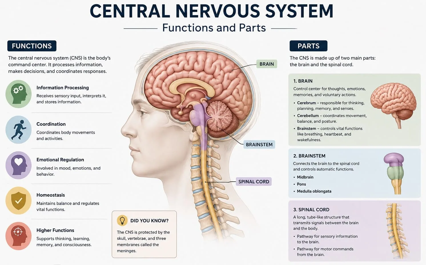
The central nervous system, often abbreviated as the CNS, is the body’s primary processing and control hub, made up of the brain and spinal cord. It serves as the supreme command center, receiving sensory information from throughout the body, integrating it with memory and ongoing needs, and generating the appropriate motor and physiological responses.
How many of these have you been "meaning to read" for months?
- 1
 HarperCollinsPete the CatI Love My White Shoes: A Groovy Story About Pete the Cat and His Colorful Shoes on a School Day A...Listen Free On Audible
 - 2
 HarvestTo Die ForA Cookbook of Gravestone RecipesListen Free On Audible
 - 3
 CrosswayDust to DustAging Wisely in an Anti-Aging WorldListen Free On Audible
The CNS carries out three broad functions that repeat constantly throughout every waking and sleeping moment. First, it receives sensory input from receptors throughout the body detecting touch, temperature, pain, sound, light, and internal chemical changes. Second, it performs integration, processing this raw information alongside stored memories, emotional context, and learned patterns to determine an appropriate response. Third, it generates motor output, sending instructions back out through the peripheral nervous system to muscles and glands, producing everything from a reflexive flinch to a deliberate, planned action.
Unlike the peripheral nervous system, which branches throughout the entire body, the central nervous system is housed entirely within the skull and vertebral column, reflecting just how critical and vulnerable this processing center truly is. This centralized location allows for coordinated, whole-body decision-making that would be impossible if processing were distributed evenly across the body’s periphery.
Because the CNS underlies virtually every physical and cognitive function, even relatively small or localized disruptions here can produce effects that feel disproportionately large, touching movement, sensation, mood, and thought all at the same time.
What Are the Main Parts of the Brain Within the CNS?
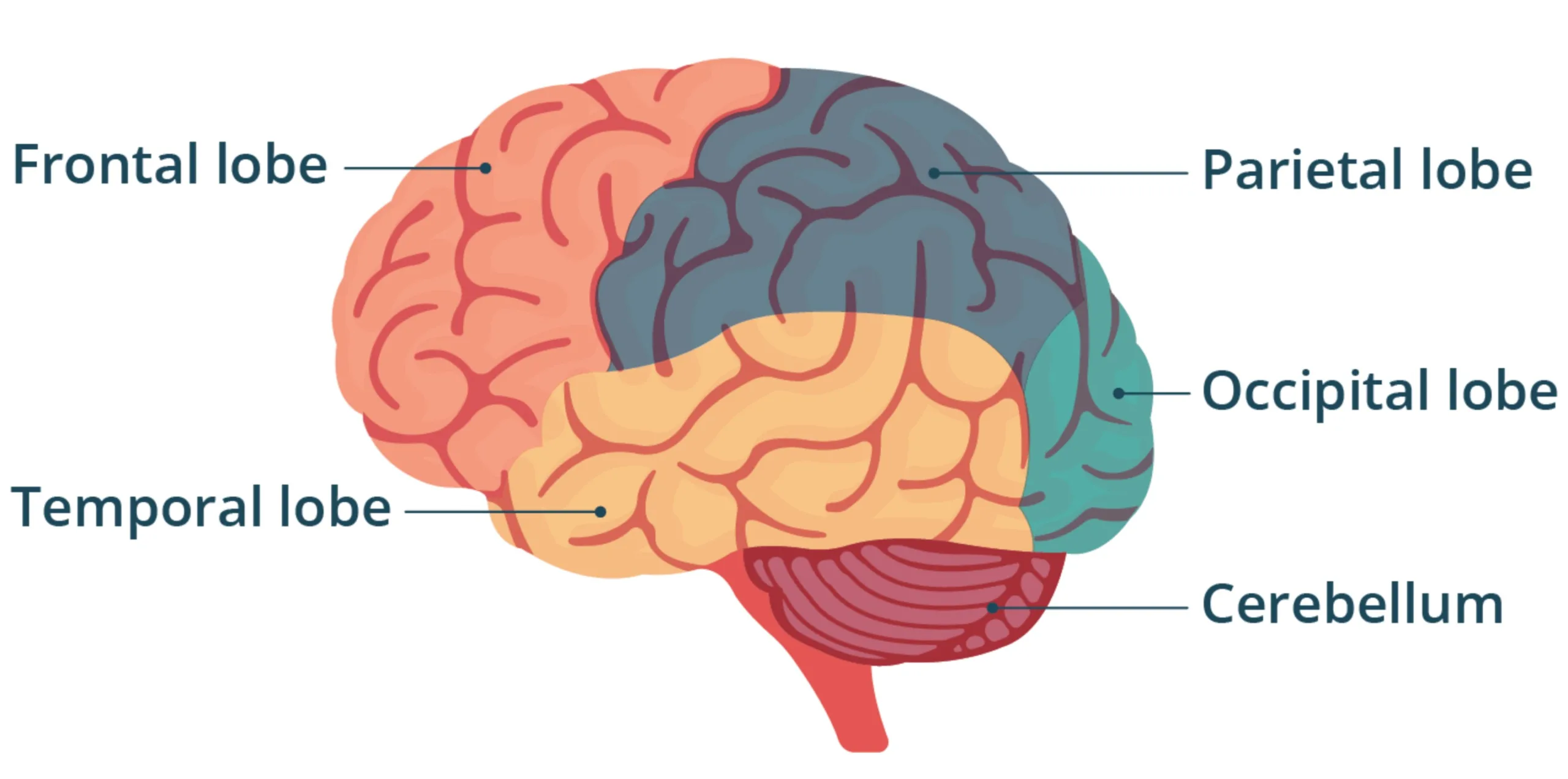
The brain is the central nervous system’s largest and most complex structure, traditionally divided into four major regions: the cerebrum, cerebellum, brainstem, and diencephalon. Each region handles a distinct category of function, though all work in close coordination.
The cerebrum, the largest portion of the brain, includes the cerebral cortex along with deeper structures like the basal ganglia, hippocampus, and amygdala, and is responsible for conscious thought, voluntary movement, language, and personality. Neurosurgeon Wilder Penfield’s pioneering work mapping the cerebral cortex through direct stimulation helped establish how specific regions of this structure control specific bodily functions, laying groundwork still used in neurology today. The cerebellum, located at the back of the brain beneath the cerebrum, coordinates movement, balance, and posture, refining motor commands to make movement smooth and precise rather than jerky or imprecise.
The brainstem, connecting the brain to the spinal cord, consists of the midbrain, pons, and medulla oblongata, and manages essential survival functions including breathing, heart rate, and blood pressure, largely without conscious control. The diencephalon, tucked deep within the brain, contains the thalamus, which relays sensory information to the cortex, and the hypothalamus, which regulates hormones and helps maintain homeostasis, a concept physiologist Walter Cannon first described as the body’s constant, automatic effort to maintain internal stability.
- The cerebrum governs conscious thought, voluntary movement, language, and personality.
- The cerebellum coordinates balance, posture, and precise motor control.
- The brainstem regulates essential survival functions like breathing and heart rate.
- The diencephalon, including the thalamus and hypothalamus, supports sensory relay and homeostasis.
How Does the Spinal Cord Function Within the Central Nervous System?
The spinal cord serves as the vital communication highway connecting the brain to the rest of the body, while also independently managing certain rapid, automatic responses. Running from the base of the brainstem down through the vertebral column, it plays a role that is both structural and functional, not merely a passive conduit.
Sensory information from the skin, muscles, and organs travels up the spinal cord toward the brain, while motor commands from the brain travel back down toward muscles and glands, all organized through distinct nerve tracts running through the spinal cord’s white matter. Physiologist Charles Sherrington’s foundational research on spinal reflexes demonstrated that the spinal cord is capable of processing certain responses entirely on its own, without waiting for input from the brain, a capacity essential for rapid protective reactions like pulling a hand away from a hot surface.
The spinal cord’s internal structure mirrors this dual role: an inner core of gray matter, containing neuron cell bodies responsible for local processing and reflexes, surrounded by an outer layer of white matter, made up of myelinated fibers carrying signals over longer distances between the spinal cord and brain. Damage at any level of the spinal cord tends to affect sensation and movement below that specific point, which is why the exact location of a spinal cord injury so precisely determines the resulting pattern of symptoms.
A practical implication of this structure: because the spinal cord processes some reflexes independently, certain automatic protective responses can remain intact even after a brain injury, offering both diagnostic clues and, in some cases, meaningful functional capacity during recovery.
How Is the Central Nervous System Protected?
The central nervous system is safeguarded by multiple layers of physical and biochemical protection, reflecting how critical and irreplaceable this tissue truly is. This protection operates on several levels simultaneously.
The skull and vertebral column provide the outermost layer of bony protection, shielding the brain and spinal cord from direct physical trauma. Beneath this bony shell, three layers of connective tissue called the meninges — the dura mater, arachnoid mater, and pia mater — wrap around the brain and spinal cord, providing additional cushioning and structural support. Between these membrane layers circulates cerebrospinal fluid, a clear liquid that cushions the CNS against sudden movement, helps remove metabolic waste, and maintains a stable chemical environment for delicate neural tissue.
An additional, less visible layer of protection comes from the blood-brain barrier, a selective filtering system formed by specialized cells lining the brain’s blood vessels, which tightly regulates what substances can pass from the bloodstream into brain tissue. This barrier helps protect the brain from toxins and pathogens circulating in the blood, though it also complicates the delivery of certain medications intended to treat brain-related conditions, a genuine challenge in modern neurological and psychiatric treatment.
Understanding these protective layers highlights why head and spinal injuries are treated with such urgency in emergency medicine; even when external protection seems adequate, forces strong enough to disrupt these barriers can produce serious, sometimes irreversible damage to the tissue they’re designed to guard.
How Does the CNS Differ From the Peripheral Nervous System?
The central and peripheral nervous systems work together as one continuous network, but they differ significantly in structure, location, and capacity for repair. Understanding this distinction clarifies why certain injuries carry very different prognoses depending on which system is affected.
| Feature | Central vs Peripheral Nervous System |
| Location | CNS: brain and spinal cord; PNS: nerves outside the CNS |
| Protection | CNS: skull, vertebrae, meninges; PNS: less structurally protected |
| Repair capacity | CNS: very limited regeneration; PNS: greater capacity for repair |
The peripheral nervous system connects the CNS to the rest of the body, carrying sensory information inward and motor commands outward, and further divides into the somatic nervous system for voluntary movement and the autonomic nervous system for involuntary functions like heart rate and digestion. One especially important practical difference lies in regenerative capacity: peripheral nerves can sometimes regrow and reestablish function after injury, while neurons within the central nervous system have a much more limited ability to regenerate, which is part of why brain and spinal cord injuries often carry a more guarded long-term prognosis than comparable peripheral nerve injuries.
This distinction matters enormously for patients and families navigating a nervous system injury, since understanding whether damage involves the central or peripheral system helps set realistic expectations for the recovery process and the types of rehabilitation likely to be most beneficial.
How Does the Central Nervous System Process Sensory Information and Movement?
The CNS continuously receives sensory input and transforms it into coordinated movement through a tightly organized processing chain. This chain begins the moment a sensory receptor detects a stimulus and ends when a muscle contracts in response.
Sensory information travels from receptors throughout the body through peripheral nerves into the spinal cord and up to the thalamus, which relays it to the appropriate region of the cerebral cortex for conscious interpretation. From there, the cortex, cerebellum, and basal ganglia work together to plan and refine an appropriate motor response, which then travels back down through the spinal cord and out to the relevant muscles. This entire cycle, from sensation to action, can happen within a fraction of a second for simple tasks, though more complex decisions naturally take longer as the brain weighs additional context and options.
The cerebellum plays a particularly important role in this process, continuously comparing intended movement with actual movement and making rapid corrections to keep motions smooth and accurate, which is why cerebellar damage often produces movements that look uncoordinated or overshoot their target despite adequate muscle strength. This fine-tuning happens largely below the level of conscious awareness, which is part of why we rarely think consciously about the mechanics of walking or reaching for an object.
A practical takeaway: because this sensory-motor processing chain depends on multiple CNS regions working together seamlessly, activities requiring coordinated movement and balance, like specific balance and coordination exercises recommended in physical therapy, can meaningfully support recovery after certain types of CNS injury by encouraging these circuits to adapt and reorganize.

How Does the CNS Support Memory, Language, and Cognition?
Beyond basic sensory and motor processing, the central nervous system underlies the full range of higher cognitive functions that define human experience, including memory, language, reasoning, and emotional regulation. These functions depend on specialized, interconnected networks distributed across the brain.
Neuropsychologist Brenda Milner’s extensive research, including landmark studies of patients with damage to the medial temporal lobe, helped establish that structures like the hippocampus are essential for forming new long-term memories, while other memory systems, such as those supporting learned motor skills, depend on entirely separate neural circuits. Language similarly relies on specialized regions identified by physician Paul Broca and neurologist Carl Wernicke in the nineteenth century, whose namesake areas in the frontal and temporal lobes respectively support language production and comprehension. More recently, neuroscientist Eric Kandel’s Nobel Prize-winning research on the cellular basis of learning and memory demonstrated that these cognitive processes depend on measurable, lasting changes in the strength of connections between neurons, a phenomenon called synaptic plasticity.
This distributed organization explains why cognitive symptoms following brain injury or disease can look so different from person to person, depending on precisely which networks are affected. A stroke affecting the language-dominant hemisphere, for instance, may leave memory and reasoning largely intact while significantly disrupting the ability to speak or understand language.
Recognizing that specific cognitive functions map to specific, identifiable brain networks can help families make sense of seemingly puzzling patterns of strength and difficulty following a CNS injury, replacing confusion with a clearer, more compassionate understanding of what recovery might realistically involve.
What Happens When the Central Nervous System Is Damaged?
CNS damage can produce a remarkably wide range of effects, depending on which specific structures are involved and the extent of the injury. Common causes include stroke, traumatic brain or spinal cord injury, tumors, infections, and neurodegenerative diseases.
Damage to different CNS regions tends to produce fairly predictable symptom patterns: cerebral injuries often affect movement, sensation, language, or personality depending on the specific lobe involved; cerebellar injuries typically cause coordination problems and unsteady movement; brainstem injuries can disrupt basic survival functions like breathing and heart rate, making them particularly serious; and spinal cord injuries produce sensory and motor loss below the level of the injury. Because CNS neurons have such limited capacity for regeneration compared to peripheral nerves, recovery from central nervous system injury tends to rely heavily on neuroplasticity, the brain’s ability to reorganize and adapt existing neural pathways, rather than actual regrowth of damaged tissue.
- Cerebral injury can affect movement, sensation, language, or personality depending on the lobe involved.
- Cerebellar damage typically produces coordination difficulties and unsteady, imprecise movement.
- Brainstem injury can disrupt essential survival functions, making it a medical emergency.
- Spinal cord injury produces sensory and motor loss below the specific level of damage.
Because CNS symptoms can appear in such varied combinations, a thorough neurological evaluation is essential for accurately identifying the location and extent of injury, guiding both immediate treatment decisions and long-term rehabilitation planning.

How Are CNS Disorders Diagnosed and Treated?
Diagnosing central nervous system disorders typically combines a detailed neurological examination with targeted imaging and, when appropriate, laboratory testing. This layered approach helps pinpoint both the location and underlying cause of dysfunction.
MRI and CT imaging remain the primary tools for visualizing structural abnormalities like stroke, tumor, or traumatic injury within the brain and spinal cord, while electroencephalography can detect abnormal electrical activity associated with seizures or other neurological conditions. Lumbar puncture, which samples cerebrospinal fluid, can help identify infections, inflammation, or certain markers associated with neurodegenerative disease. A thorough neurological exam, assessing reflexes, strength, sensation, coordination, and cognition, often provides crucial localizing information even before imaging results are available.
Treatment depends entirely on the underlying cause and can range from medication and physical rehabilitation to surgical intervention for structural problems like tumors or severe traumatic injury. Rehabilitation following CNS injury often combines physical therapy, occupational therapy, and speech-language therapy, tailored to the specific pattern of deficits, with early, consistent engagement generally supporting better long-term functional outcomes.
- Seek immediate emergency care for any sudden weakness, confusion, severe headache, or loss of coordination, since rapid treatment can significantly affect outcomes in conditions like stroke.
- Request a thorough neurological evaluation for persistent, unexplained cognitive or physical symptoms rather than assuming they will resolve on their own.
- Engage consistently in rehabilitation following a CNS injury, since neuroplasticity supports meaningful recovery with sustained, structured practice.
- Follow up on cerebrospinal fluid or imaging findings promptly, since early diagnosis often allows for more effective, less invasive treatment.
How Does the Central Nervous System Change Across the Lifespan?
The central nervous system develops rapidly before birth and continues maturing well into early adulthood, while also undergoing predictable changes in older age. Recognizing this trajectory helps distinguish typical development and aging from patterns that warrant closer medical evaluation.
In early childhood, the CNS undergoes extraordinary neuron growth followed by extensive synaptic pruning and myelination, processes that refine neural connections and dramatically improve processing speed and coordination as children develop increasingly complex motor and cognitive skills. This maturation continues into adolescence and early adulthood, particularly within the prefrontal regions of the cerebrum responsible for executive function, judgment, and impulse control, which explains why these capacities continue developing well beyond childhood. In older adulthood, gradual changes in brain volume and processing speed are a normal part of aging, though certain neurodegenerative conditions, including Alzheimer’s disease and Parkinson’s disease, involve more significant and progressive CNS changes that go well beyond typical age-related decline.
Supporting central nervous system health across the lifespan includes managing cardiovascular risk factors that affect blood flow to the brain and spinal cord, protecting against head and spinal injuries, staying physically and cognitively active, and prioritizing consistent sleep, all of which have been associated with better-preserved CNS structure and function over time.
Recognizing that some degree of CNS change is a normal part of aging, distinct from a more serious progressive condition, can help ease unnecessary worry while still supporting timely medical evaluation for any sudden or rapidly progressing symptoms.
FAQs about the Central Nervous System
What is the main function of the central nervous system?
The central nervous system’s core function is to receive sensory information, integrate and process it, and generate an appropriate motor or physiological response. It coordinates virtually every aspect of thought, movement, sensation, and involuntary bodily function, acting as the body’s supreme processing and command center. Because the brain and spinal cord work together so closely, damage to either structure can affect a wide range of physical and cognitive abilities. This central role is why the CNS is considered the most critical and closely protected part of the entire nervous system.
What are the two main parts of the central nervous system?
The central nervous system consists of two main structures: the brain and the spinal cord. The brain further divides into the cerebrum, cerebellum, brainstem, and diencephalon, each responsible for distinct functions ranging from conscious thought to basic survival regulation. The spinal cord serves as the primary communication pathway between the brain and the rest of the body, while also independently managing certain automatic reflexes. Together, these two structures are protected by the skull, vertebral column, meninges, and cerebrospinal fluid, reflecting how essential and vulnerable this tissue truly is.
How is the central nervous system different from the peripheral nervous system?
The central nervous system consists of the brain and spinal cord, while the peripheral nervous system includes all the nerves branching out from that central hub to the rest of the body. The CNS is more heavily protected by bone and connective tissue and has a much more limited capacity for repair after injury, while peripheral nerves generally have somewhat greater regenerative potential. The peripheral nervous system further divides into the somatic nervous system, controlling voluntary movement, and the autonomic nervous system, managing involuntary functions like heart rate and digestion. Both systems work together continuously, even though they differ significantly in structure and injury recovery potential.
What happens if the central nervous system is damaged?
Symptoms of central nervous system damage vary widely depending on which specific structures are affected. Cerebral injury can disrupt movement, sensation, language, or personality; cerebellar damage typically causes coordination and balance problems; brainstem injury can threaten essential survival functions like breathing; and spinal cord injury produces sensory and motor loss below the level of the injury. Because CNS tissue has limited capacity for regeneration, recovery often relies heavily on neuroplasticity and structured rehabilitation rather than actual tissue regrowth. A thorough neurological evaluation is essential for understanding the specific pattern and extent of any CNS injury.
Can the central nervous system heal itself after injury?
The central nervous system has a much more limited capacity for self-repair compared to the peripheral nervous system, since neurons within the brain and spinal cord generally do not regenerate the way peripheral nerves sometimes can. However, the brain retains a genuine capacity for neuroplasticity, allowing surviving neural pathways to reorganize and take over some functions previously handled by damaged tissue. This adaptive capacity underlies much of the meaningful functional recovery seen after stroke, traumatic brain injury, or spinal cord injury, particularly with consistent, structured rehabilitation. Recovery potential varies significantly depending on the severity, location, and cause of the injury, along with the individual’s age and overall health.
What are common signs of a central nervous system disorder?
Common signs of central nervous system dysfunction include persistent headaches, unexplained weakness or numbness, coordination or balance difficulties, changes in vision or speech, memory or cognitive changes, and seizures. Because the CNS controls such a wide range of functions, symptoms can appear quite varied and sometimes seem unrelated to one another at first glance. Sudden, severe symptoms like facial drooping, slurred speech, or sudden weakness on one side of the body require immediate emergency evaluation, since they can indicate a stroke or other time-sensitive neurological emergency. Persistent or gradually worsening symptoms should also be evaluated promptly by a healthcare provider rather than assumed to be temporary.
Bibliography
- StatPearls Publishing. (2026). Anatomy, Central Nervous System. National Center for Biotechnology Information Bookshelf.
- Cleveland Clinic. (2023). Central Nervous System (CNS): What It Is & Function.
- Kenhub. (2015). Central Nervous System: Anatomy, Structure, Function.
- Penfield, W., & Rasmussen, T. (1950). The Cerebral Cortex of Man: A Clinical Study of Localization of Function. Macmillan.
- Sherrington, C. S. (1906). The Integrative Action of the Nervous System. Yale University Press.
- Cannon, W. B. (1932). The Wisdom of the Body. W.W. Norton & Company.
- Broca, P. (1861). Remarks on the seat of the faculty of articulate language, followed by an observation of aphemia. Bulletin de la Société Anatomique de Paris.
- Wernicke, C. (1874). Der aphasische Symptomencomplex. Cohn and Weigert.
- Milner, B., Corkin, S., & Teuber, H. L. (1968). Further analysis of the hippocampal amnesic syndrome. Neuropsychologia.
- Kandel, E. R. (2006). In Search of Memory: The Emergence of a New Science of Mind. W.W. Norton & Company.
Use this citation format to reference the article clearly and help readers find the original source.
PsychologyFor. (2026). Central Nervous System: Functions and Parts. PsychologyFor. https://psychologyfor.com/central-nervous-system-functions-and-parts/