Cerebral Angiography: What Is It And What Disorders Can It Detect?
All our organs require the action of the cardiovascular system to survive, because thanks to it they receive the oxygen and nutrients necessary for their survival and proper functioning.
This includes our nervous system, which requires a continuous supply of these elements. But sometimes there may be alterations that damage the vascular system that irrigates the brain or symptoms that suggest the existence of said damage.
For this reason, it is necessary to have different techniques that allow us to observe and analyze the blood flow of the brain, being one of the best known cerebral angiography
Cerebral angiography is a medical evaluation technique that allows the study and analysis of cerebral flow and the health of the cerebrovascular system. This is a technique in which X-rays are used to visualize by injecting contrast into the main brain blood vessels the flow and state of the circulatory system. The images obtained are generally very clear and allow precise identification of alterations in the blood circulation of the brain.
The procedure is as follows: after placing the patient on the X-ray table, the head is immobilized and a sedative is administered while cardiac activity is monitored. After this, the patient is inserted a catheter into the arteries of the arm or leg, which will be guided through the artery to the neck with the help of X-rays. Once there, a contrast solution is injected through the catheter. to later take images of the blood circulation using x-rays. After this, and unless some type of intervention has to be carried out through it, the catheter is removed and pressure is applied to the area through which it has been introduced in order to avoid bleeding.
Although it is generally used as a technique for the diagnosis and monitoring of cerebrovascular disorders, the fact that a catheter is used to perform it allows therapeutic procedures such as the delivery of drugs to be used in addition, which can prevent need for other treatments.
Guys
Cerebral angiography is a technique that has various variants depending on the mechanisms used to evaluate the state of the patient’s blood vessels. Some of the best known are the following
1. Conventional angiography (by intra-arterial digital subtraction)
This is the previously explained procedure, in which the catheter is placed in the artery and guided to its objective. This is an invasive procedure that is usually the most common due to its effectiveness and the high level of clarity it allows. The catheter is usually introduced femorally, through the groin to the aortic arch, where after a first injection of contrast, the catheter is placed in the artery that needs to be analyzed.
Regarding digital subtraction, it refers to the fact that frequently in x-rays the skull is digitally removed from the image taken, so that the image of the blood vessels can be observed more clearly.
2. Helical computed tomography angiography
In this case, no type of catheter is introduced into the subject’s body, but it does require the injection of contrast in order to obtain the image using CT. It is less invasive than its conventional counterpart.
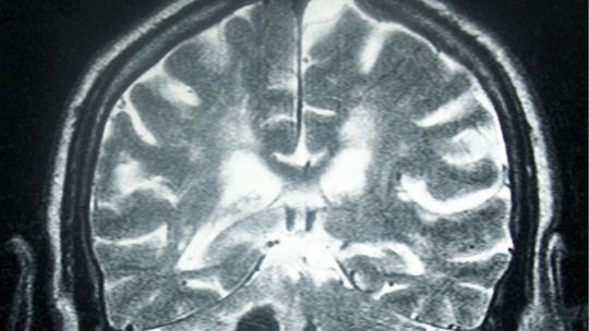
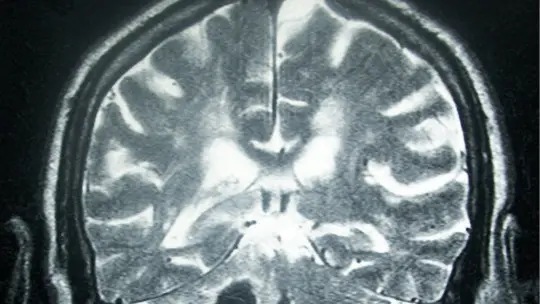
3. Magnetic resonance angiography
In this type of angiography, no catheter is used either, as it is not an invasive technique. It involves performing an MRI, not using radiation as in other cases.
Cerebral angiography is a test that even today It is used as one of the main ones to observe the circulatory flow and the state of the blood vessels of the brain There are multiple disorders and diseases that the application of this technique allows us to observe.
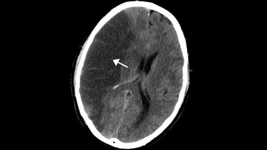
1. Cerebrovascular accidents or stroke
Angiography allows us to observe the existence of extravasation and ruptures of blood vessels, or the absence or obstruction of circulation in some area of the brain. It is because of that This is a valid technique both to detect ischemia and to visualize cerebral hemorrhages (More information about strokes).
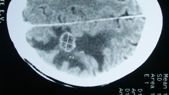
2. Aneurysms
The use of angiography allows detecting the presence of aneurysms , relatively weaker-walled, blood-filled bulges in the arterial wall that can rupture. (More information about aneurysms).
3. Tumors
The presence of tumors in the brain tends to cause alterations in blood flow to the brain , as well as causing phenomena such as strokes. For this reason, angiography allows us to observe the presence of abnormalities generated by the presence of tumors. (More information about brain tumors).
4. Malformations
The existence of congenital malformations, as occurs in AVM, can also be assessed using this evaluation and diagnosis technique.
5. Arterial or venous alterations
Through cerebral angiography, it can be observed if the blood vessels in the brain are in good health, if they are inflamed, or if disorders such as atherosclerosis occur.
6. Brain death
Cerebral angiography is also used to evaluate whether or not brain death exists. Specifically, it evaluates whether or not there is blood flow, observing an absence of irrigation in cases of brain death.
There is the possibility of observing, through cerebral angiography, the presence of different disorders and diseases apart from those mentioned above. For example, alterations may be found in neurosyphilis, or in people with disorders such as Kleine-Levine syndrome.
Risks and possible side effects of this technique
Cerebral angiography is a generally safe technique and does not tend to cause complications but this does not prevent it from having risks and adverse side effects that can cause alterations of variable severity.
One of the risks is the possibility that the patient may be allergic to the contrast applied (generally iodinated). Likewise, this could cause discomfort or even destruction of some tissues if it extravasates outside the vein. It can also be risky or harmful for people with kidney problems or diabetes.
The existence of symptoms such as tingling, breathing difficulties, vision problems, infection of the route through which the catheter has entered, problems with control of the limb into which it has been inserted, speech problems or hemiparesis are a sign that There may be some type of complication that needs to be treated quickly.
Finally, special caution is necessary in the case of pregnant or breastfeeding women, since the radiation emitted could be harmful. It may also occur that the artery is torn, generating some type of bleeding or clots that can block the vessel, although this is very rare.