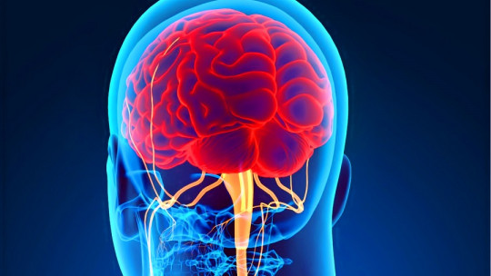
Cochlea: What It Is, Parts, Functions And Associated Pathologies
Hearing, as its name indicates, is a term that encompasses the physiological processes that give human beings the ability to hear and relate to their environment based on this essential sense.
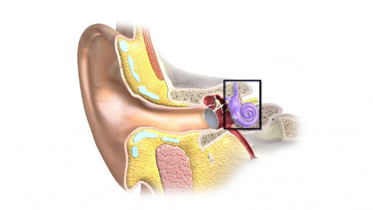
In very general terms, the hearing process can be distinguished in the following events: the ear receives sound waves, which are transmitted through the ear canal to the eardrum, which produces a series of vibrations. These reach the chain of ossicles, responsible for transmitting them to the inner ear through the oval window.
This is where it comes into play. the cochlea or snail, an essential part of the mammalian auditory system Immerse yourself with us in the world of auditory anatomy, because today we tell you what the cochlea is, its parts, the functions it performs and what happens when it fails.
The cochlea is a spirally coiled tube-shaped structure located in the inner ear, more specifically, in the temporal bone In general, this structure is about 34 millimeters long in an adult individual and, it should be noted, that the organ of Corti is located inside.
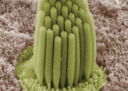
The organ of Corti is essential to understanding the hearing process, as it is made up of a series of sensory cells (approximately 16,000) arranged in a row, specifically called “hair cells.” These are the last ones in charge of “interpreting” the sound waves received by the external ear, as they transform them into electrical impulses that reach the auditory nerve, and from there, to the brain.
Parts of the cochlea
It is not yet time to describe the complex process that involves the integration of sounds at the brain level, since we still have a lot of fabric to cut in an anatomical field. In the first instance, we can say that The cochlea is made up of three essential parts We describe each of them:
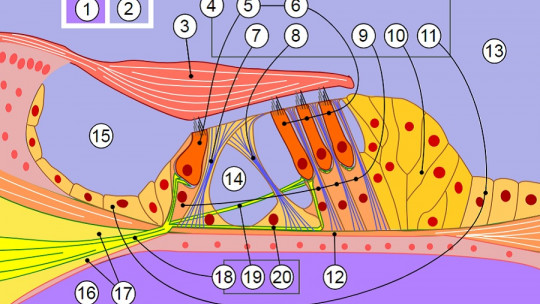
It should be noted that, beyond a description of the tissues observed in a structural cross section, it gives us more information to look at the three longitudinal chambers that make up the cochlea These are the following:
The scala tympani and scala vestibuli contain perilymph (a fluid similar to serum) and communicate with each other through a small duct called the helicotrema, located at the end of the cochlea. This allows communication and perilymph fluid between both structures. For its part, the scala media or cochlear duct is located between the scala vestibularis and the scala tympani and contains the endolymph. This structure has a fairly complex anatomy in terms of terminology, which is why we will limit ourselves to saying that it is triangular and that, finally, between the scala tympani and the scala media is the already named organ of Corti.
Beyond this conglomerate, we must also highlight that these three chambers (scala tympani, vestibular and middle) They are separated by two types of membrane: the Reissner membrane and the basilar membrane
Reissner’s membrane separates the vestibular scala and the media, and its function is to keep the endolymph in the cochlear duct, where it should remain. On the other hand, the basilar membrane is responsible for separating the scala media and scala tympani. Its function, however, is not so simple to explain, since the organ of Corti rests on it. Let’s focus a little more on this very special membrane.
The role of the basilar membrane in hearing
First of all, it is necessary to highlight that The response of the basilar membrane to certain sounds will be affected by its mechanical properties which vary progressively from the base to the apex.
At the end closest to the oval window and the eardrum, this membrane has a more rigid, thick and narrow morphology. Therefore, its resonance frequency is high for high tones. On the other hand, at the distal end the basilar membrane is wider, softer and more flexible, which causes the response to be better at low frequencies. As a curious fact, we can say that this structure produces a ten thousand-fold decrease in its rigidity from the proximal to the distal end.
At each point of this special membrane a tuning occurs , and the place where the greatest displacement occurs at a given frequency is called the “characteristic frequency”. In other words, the range of resonance frequencies available in the basement membrane determines the hearing capacity of the human being, which is between 20 Hz-20,000 Hz.
The organ of Corti
The basilar membrane analyzes the frequencies, but it is the organ of Corti is responsible for decoding this information and sending it to the brain Let’s start from the beginning to understand how it works.
We return to the base of the inner ear: when a vibration is transmitted through the ossicles of the middle ear to the oval window, a pressure difference is produced between the vestibular and tympanic cochlear scalas. Consequently, the endolymph present in the scala media moves, producing a traveling wave that propagates along the basilar membrane.
The displacements of the basilar membrane cause the hair cells (remember that they are the ones that make up the organ of Corti) to move in relation to it and, thanks to this, they are excited or inhibited depending on the direction of movement. Depending on the region of the basilar membrane that oscillates with greater amplitude according to the perceived sound, different portions of the hair cells that make up the organ of Corti will be activated.
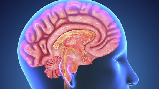
Finally, hair cells produce certain chemical components that are translated into nerve signals, which will be sent first to the acoustic nerve and then to the auditory nerve (also known as cranial nerve VIII). Of course, we are facing a journey of very complex understanding, but we can summarize it in the following concept: the basilar membrane “vibrates” more at one point or another depending on the type of sound, and the excited cells translate this signal, which ends up arriving to the brain through a series of nerves.
What happens when the cochlea fails?
Notably hair cells do not regenerate That is, when they are injured in an individual, he or she loses hearing irreparably. Human beings take our senses for granted until we lose them and, therefore, the World Health Organization (WHO) helps us contextualize a little what hearing loss means at a general level:
An important factor that promotes hearing loss (hearing loss) is chronic exposure to loud sounds In these cases, the hair cells already described or the nerves that supply them are damaged at some point, which leads the patient to hear the sound in a distorted way or, for example, to have an easier time interpreting some frequencies than others..
Finally, it is also essential to highlight that hearing loss due to age (presbycusis) is, unfortunately, completely normal. This process It is observed in almost 80% of elderly people over 75 years of age and is caused by a deterioration of the structures located in the inner ear or the auditory nerve itself.
Summary
As we have seen in these lines, the cochlea had many more secrets for us than we could imagine. From a complex morphology to the basilar membrane and the organ of Corti, one concept is clear: hearing is a true work of engineering. Maybe all this information will make us think twice the next time we turn the headphone volume up to maximum, right?
What is the cochlea? Audifon, hearing centers. Collected on November 12 at https://audifon.es/que-es/c/coclea-o-caracol/
Hearing and the cochlea, Medlineplus.gov. Collected on November 12 at https://medlineplus.gov/spanish/ency/anatomyvideos/000063.htm
Cochlea, generalities: journey into the world of hearing, cochlea.eu. Collected on November 12 at http://www.cochlea.eu/es/coclea
Cochlea, vestib.org. Collected on November 12 at https://www.vestib.org/es/coclea.html
Deafness, World Health Organization (WHO). Collected on November 12 at https://www.who.int/es/news-room/fact-sheets/detail/deafness-and-hearing-loss
Soto, E., Vega, R., Chávez, H., & Ortega, A. (2003). Physiology of hearing: the cochlea. Autonomous University of Puebla. Retrieved from: http://www. physiology. boop. mx/online/DrSotoE/COCLEA, 202003.
Terreros, G., Wipe, B., León, A., & Délano, PH (2013). From the auditory cortex to the cochlea: Progress in the auditory efferent system. Journal of Otolaryngology and Head and Neck Surgery, 73(2), 174-188.