Ion Channels: What They Are, Types. And How They Work In Cells
Ion channels are protein complexes located in cell membranes, which regulate vital processes such as heartbeat or the transmission of signals between neurons.
In this article we are going to explain what they consist of, what their function and structure is, what types of ion channels exist and their relationship with various diseases.
We understand ion channels protein complexes full of aqueous pores, which allow ions to pass , causing them to flow from one side of the cell membrane to the other. These channels are present in all cells, of which they are an essential component.
Each cell is surrounded by a membrane that separates it from the outside environment. Its lipid bilayer structure is not easily permeable to polar molecules such as amino acids or ions. Therefore, it is necessary to transport these substances in and out of the cell using membrane proteins such as pumps, transporters and ion channels.
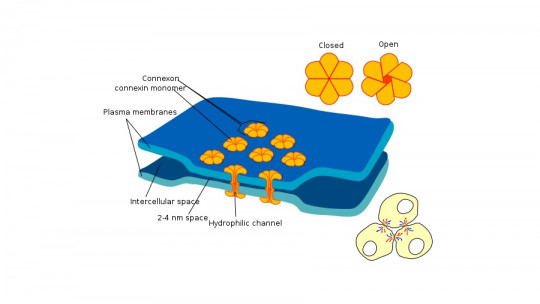
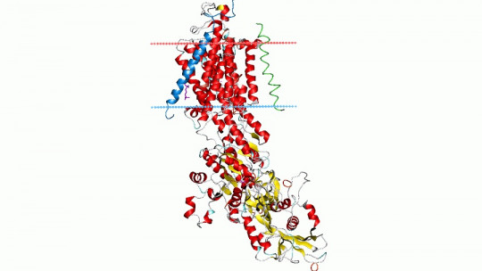
The channels They are made up of one or several different proteins called subunits (alpha, beta, gamma, etc.). When several of them come together, they create a circular structure in the center of which there is a hole or pore, which allows the passage of ions.
One of the peculiarities of these channels is their selectivity; that is, they determine that some inorganic ions pass and not others depending on the diameter and distribution of its amino acids.
The opening and closing of ion channels is regulated by various factors; A specific stimulus or sensor is what determines that they fluctuate from one state to another by altering their composition.
Let’s now see what functions they perform and what their structure is.
Functions and structure
Behind essential cellular processes, such as the secretion of neurotransmitters or the transmission of electrical signals, are ion channels, which confer electrical and excitable capacities to cells And when they fail, numerous pathologies can occur (which we will talk about later).
The structure of ion channels is in the form of transmembrane proteins and They act like a system of gates to regulate the passage of ions (potassium, sodium, calcium, chlorine, etc.) through pores.
Until a few years ago, it was thought that the pores and the voltage sensor were coupled through a linker (a spiral of about 15 amino acids), which can be activated by the movement of the voltage sensor. This coupling mechanism between the two parts of the ion channel is the canonical mechanism that has always been theorized.
However, recently, new research has revealed another way that involves a segment of amino acids constituted by part of the voltage sensor and part of the pore These two segments would adjust like a kind of zipper to trigger the opening or closing of the channel. In turn, this new mechanism could explain recent discoveries, in which some voltage-regulated ion channels (some responsible for functions such as heartbeat) with just one linker have been detected.
Voltage-gated ion channels are only one of the existing types of channels, but there are more: let’s see what they are below.
Types of ion channels
The mechanisms for the activation of ion channels can be of several types: by ligand, by voltage or by mechanosensitive stimuli.
1. Ligand-gated ion channels
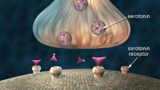
These ion channels They open in response to the binding of certain molecules and neurotransmitters This opening mechanism is due to the interaction of a chemical substance (which can be a hormone, a peptide or a neurotransmitter) with a part of the channel called a receptor, which generates a change in free energy and modifies the conformation of the protein by opening the channel.
The nicotinic-type acetylcholine receptor (a neurotransmitter involved in the transmission of signals between motor nerves and muscles) is one of the most studied ligand-gated ion channels. It is composed of 5 subunits of 20 amino acids and is involved in basic functions such as the voluntary control of movement, memory, attention, sleep, alertness, or anxiety
This type of channels open in response to changes in electrical potential across the plasma membrane Voltage-gated ion channels are involved in the transmission of electrical impulses, generating action potentials due to changes in the difference in electrical charges on both sides of the membrane.
The flow of ions is carried out in two processes: by activation, a voltage-dependent process: the channel opens in response to changes in the membrane potential (electrical potential difference on both sides of the membrane); and inactivation, a process that regulates channel closure.
The main function of voltage-gated ion channels is the generation of action potentials and their propagation There are several types and the main ones are:
2.1. Na+ channel
They are transmembrane proteins that allow the passage of sodium ions through the cell. Ion transport is passive and only depends on the electrochemical potential of the ion (it does not require energy in the form of an ATP molecule). In neurons, sodium channels are responsible for the ascending phase of the action potential (depolarization).
2.2. K+ channel
These ion channels constitute the most heterogeneous group of structural membrane proteins. In neurons, depolarization activates K+ channels and facilitates K+ efflux from the nerve cell, leading to a repolarization of the membrane potential.
23. Ca++ channel
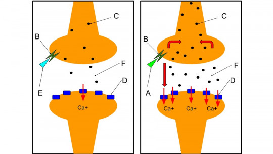
Calcium ions promote the fusion of the membrane of the synaptic vesicle (structures located at the end of the neuronal axon and responsible for secreting neurotransmitters) with the terminal membrane of the axon in the neuron, stimulating the release of acetylcholine into the synaptic cleft by an exocytosis mechanism
2.4. Cl- channel
This type of ion channels are responsible for regulating cell excitability, transport between cells, as well as the management of PH and cell volume. Channels located in the membrane stabilize the membrane potential in excitable cells. They are also responsible for transporting water and electrolytes between cells
3. Ion channels regulated by mechanosensitive stimuli
These ion channels open in response to mechanical actions They can be found, for example, in Paccini corpuscles (sensory receptors in the skin that respond to rapid vibrations and deep mechanical pressure), which open by stretching the cell membrane through the application of tension and/or pressure.
Channelopathies: pathologies associated with these molecules
From a physiological point of view, ion channels They are essential for the homeostatic balance of our body Its dysfunction causes a whole series of diseases, known as channelopathies. These can be produced by two types of mechanisms: genetic alterations and autoimmune diseases.
Among the genetic alterations, there are mutations that occur in the coding region of the gene for an ion channel. It is common for these mutations to produce polypeptide chains that are not processed correctly and are not incorporated into the plasma membrane; or, when the subunits couple and form the channels, they are not functional.
Another common possibility is that, even though they are functional channels, they end up showing altered kinetics. Whatever the case, they usually lead to the gain or loss of the channel’s function.
Also mutations can occur in the promoter region of the gene that codes for an ion channel This can cause underexpression or overexpression of the protein, producing changes in the number of channels, which would also cause an increase or decrease in its functionality.
Currently, multiple pathologies associated with ion channels in different tissues are known. At the musculoskeletal level, mutations in voltage-gated Na+, K+, Ca++ and Cl- channels and in the acetylcholine channel lead to disorders such as hyper and hypokalemic paralysis, myotonia, malignant hyperthermia and myasthenia
At the neuronal level, it has been proposed that alterations in voltage-activated Na+ channels, voltage-activated K+ and Ca++ channels, the acetylcholine-activated channel or the glycine-activated channel could explain disorders such as epilepsy, episodic ataxia , familial hemiplegic migraine, Lambert-Eaton syndrome, Alzheimer’s disease, Parkinson’s disease and schizophrenia.