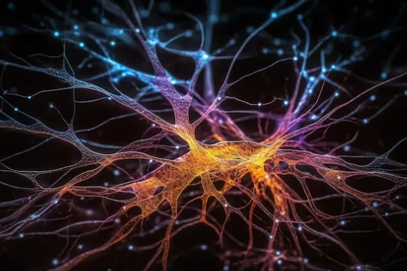
Your brain is having about 100 trillion conversations right now. Neurons are firing, chemicals are flooding across microscopic gaps, and somewhere in that chaos, you’re reading these words, understanding them, maybe feeling curious or skeptical or bored. None of this would be possible without neural receptors—the molecular machinery that allows one neuron to tell another neuron what to do. These specialized proteins sit on neuron surfaces like locks waiting for the right chemical keys, and when the right neurotransmitter binds to them, everything from your heartbeat to your deepest thoughts depends on what happens next.
Think of neural receptors as the nervous system’s translators. A neurotransmitter molecule—dopamine, serotonin, glutamate, GABA—carries a message across the synaptic gap between neurons. But that chemical message means nothing until it binds to a receptor on the receiving neuron that can interpret it and translate it into electrical or biochemical signals that the neuron understands. Without receptors, neurotransmitters would just float around uselessly, like messages in bottles that nobody can open.
What makes this system fascinating is its specificity. Each receptor only responds to particular neurotransmitters, like locks accepting only certain keys. The same neurotransmitter can have completely different effects depending on which receptor it activates. Acetylcholine binding to nicotinic receptors causes rapid muscle contraction. The same acetylcholine binding to muscarinic receptors produces slower, longer-lasting effects on heart rate and digestion. One chemical, multiple meanings, determined entirely by which receptor receives it.
The study of neural receptors revolutionized neuroscience and pharmacology. Understanding how these proteins work explained not just normal brain function but also what goes wrong in neurological and psychiatric disorders. Depression, schizophrenia, Parkinson’s disease, Alzheimer’s, addiction—all involve receptor dysfunction or imbalances in the neurotransmitter systems that activate them. Most psychiatric medications work by targeting specific receptors—enhancing activity at some, blocking activity at others, fine-tuning the neurochemical conversations that determine mood, cognition, and behavior.
This article explores neural receptors comprehensively: what they are at the molecular level, the two major types and how they differ, the specific receptors for major neurotransmitters, how receptors translate chemical signals into cellular responses, what happens when receptor systems malfunction, and why understanding these molecular gatekeepers matters for everything from developing better medications to understanding consciousness itself.
What Neural Receptors Actually Are
Neural receptors, more precisely called neurotransmitter receptors, are specialized protein molecules embedded in neuron membranes that detect and respond to chemical signals. They’re the receiving end of neural communication—when one neuron releases neurotransmitter molecules into the synaptic cleft (the tiny gap between neurons), receptors on the receiving neuron bind to those molecules and initiate responses inside the target cell.
These aren’t just simple on-off switches but sophisticated molecular machines with specific shapes that determine which chemicals can bind to them and what happens when binding occurs. The receptor protein spans the cell membrane, with parts extending into the space outside the cell (where they can bind neurotransmitters), parts embedded within the membrane itself, and parts extending into the cell’s interior (where they trigger intracellular responses).
The lock-and-key analogy is often used, but it’s more accurate to think of receptors as shape-shifters. When a neurotransmitter binds to a receptor, it doesn’t just fit into a static pocket—the binding causes the receptor protein to change shape, and this conformational change is what triggers the cellular response. It’s like a key not just unlocking a door but the door transforming into something entirely different when unlocked.
Receptors exist in different states—resting, activated, desensitized, and inactivated. In the resting state, the receptor is available for neurotransmitter binding. Binding activates it, triggering responses. But with prolonged activation, receptors can become desensitized—they stop responding even though the neurotransmitter is still present. This desensitization prevents overstimulation and explains why some drugs become less effective with repeated use.
The specificity of receptors is remarkable but not absolute. Most receptors bind most strongly to one primary neurotransmitter but can also bind to chemically similar molecules with lower affinity. This is how drugs work—they’re designed to mimic neurotransmitters closely enough to bind receptors and either activate them (agonists) or block them without activating (antagonists). Understanding receptor structure and binding properties allowed pharmaceutical companies to design molecules that selectively target specific receptor subtypes, creating medications with fewer side effects than earlier drugs that activated many receptors indiscriminately.
The Two Major Types: Ionotropic and Metabotropic
Neural receptors divide into two fundamentally different classes based on how they translate neurotransmitter binding into cellular responses: ionotropic receptors and metabotropic receptors. The difference isn’t just academic—it determines the speed, duration, and nature of neural communication.
Ionotropic receptors are ligand-gated ion channels—the receptor and the ion channel are the same molecule. When a neurotransmitter (the ligand) binds, the receptor changes shape to open a channel through the membrane, allowing specific ions to flow in or out. This happens incredibly fast—within milliseconds—because there are no intermediary steps. The neurotransmitter binds, the channel opens, ions flow, and the electrical properties of the neuron change immediately.
Think of ionotropic receptors as direct connections. The signal (neurotransmitter binding) produces an immediate effect (ion flow and electrical change). This makes ionotropic receptors perfect for rapid communication—reflexes, fast sensory processing, immediate motor responses. When you touch something hot and jerk your hand away before consciously registering pain, that’s ionotropic receptors at work.
Metabotropic receptors work differently. They’re G-protein coupled receptors (GPCRs) that don’t have ion channels built into them. Instead, when a neurotransmitter binds, the receptor activates a G-protein inside the cell, which then triggers a cascade of intracellular events. This cascade might eventually open or close ion channels, but through indirect pathways involving multiple molecular steps. The process takes longer—hundreds of milliseconds to seconds—but produces more sustained, modulated responses that can amplify signals and affect multiple cellular processes simultaneously.
Metabotropic receptors are like indirect connections that allow for more complex signal processing. One neurotransmitter binding to one metabotropic receptor can trigger production of many second messenger molecules, which then affect multiple cellular targets. This amplification means a small amount of neurotransmitter can produce large effects. The slowness is a feature, not a bug—it allows for sustained modulation of neural activity rather than just brief on-off signals.
Many neurotransmitters can bind to both ionotropic and metabotropic receptors, producing both fast and slow effects simultaneously. Glutamate activates ionotropic receptors (AMPA and NMDA) for rapid excitation and metabotropic glutamate receptors for slower, modulatory effects. Acetylcholine activates nicotinic receptors (ionotropic) for fast signaling and muscarinic receptors (metabotropic) for slower effects. This dual system allows sophisticated control over neural communication.
Ionotropic Receptors: Fast Communication
Ionotropic receptors mediate fast synaptic transmission throughout the nervous system. They’re responsible for the rapid-fire communication that makes reflexes, sensory processing, and quick movements possible. Understanding specific ionotropic receptor types reveals how different neurotransmitters produce different effects despite using similar mechanisms.
Nicotinic acetylcholine receptors (nAChRs) are found at neuromuscular junctions where motor neurons connect to muscles, and throughout the brain. When acetylcholine binds, sodium ions rush into the cell, depolarizing the membrane and triggering action potentials. At muscles, this causes contraction—acetylcholine from motor neurons tells muscles to move. In the brain, nicotinic receptors modulate attention, learning, and memory. Nicotine from cigarettes activates these receptors, which is why smoking affects alertness and can be addictive.
AMPA receptors are the workhorses of fast excitatory transmission in the brain. They bind glutamate, the primary excitatory neurotransmitter, and allow sodium influx that depolarizes neurons. Most rapid information transfer in the brain—processing sensory information, motor commands, thinking—involves AMPA receptor activation. They’re named after a synthetic chemical (alpha-amino-3-hydroxy-5-methyl-4-isoxazolepropionic acid) that selectively activates them.
NMDA receptors are also activated by glutamate but work differently than AMPA receptors. They require both glutamate binding and membrane depolarization to open, making them coincidence detectors—they only fully activate when the presynaptic neuron releases glutamate AND the postsynaptic neuron is already somewhat depolarized. This property makes NMDA receptors crucial for synaptic plasticity, learning, and memory formation. They allow calcium influx, which triggers intracellular cascades that strengthen or weaken synaptic connections.
GABA-A receptors are the primary inhibitory ionotropic receptors in the brain. When GABA (gamma-aminobutyric acid) binds, chloride ions flow into the cell, hyperpolarizing it and making it less likely to fire action potentials. This inhibition is essential for preventing excessive neural activity—without sufficient GABA-A receptor function, neurons would fire uncontrollably, causing seizures. Benzodiazepines (like Valium) and barbiturates enhance GABA-A receptor function, producing sedation and anxiety reduction.
Glycine receptors provide inhibitory transmission primarily in the spinal cord and brainstem. Like GABA-A receptors, they allow chloride influx causing hyperpolarization. Strychnine, a poison, blocks glycine receptors, causing convulsions and death by preventing inhibitory control of motor neurons.
Metabotropic Receptors: Slow Modulation
Metabotropic receptors provide slower, longer-lasting effects that modulate neural activity rather than producing immediate electrical changes. They’re involved in everything from mood regulation to hormone release to pain perception. All metabotropic receptors are G-protein coupled receptors, but they activate different intracellular pathways producing diverse effects.
Muscarinic acetylcholine receptors (mAChRs) are found throughout the brain and in peripheral organs like the heart and digestive system. Unlike nicotinic receptors’ rapid effects, muscarinic receptors produce slower changes in neural excitability and can either increase or decrease neuronal activity depending on the subtype. M1, M3, and M5 subtypes tend to be excitatory through their effects on ion channels and intracellular signaling, while M2 and M4 subtypes are generally inhibitory. Medications for overactive bladder block muscarinic receptors. Pilocarpine, used to treat glaucoma, activates them.
Dopamine receptors are crucial for motivation, reward, movement, and cognition. There are five subtypes (D1-D5) divided into two families based on their effects. D1 and D5 receptors generally increase neural excitability by activating adenylyl cyclase, which produces cAMP (a second messenger). D2, D3, and D4 receptors generally decrease excitability by inhibiting adenylyl cyclase. Parkinson’s disease involves loss of dopamine neurons, causing movement problems that can be partially treated with dopamine precursors or dopamine agonists that activate dopamine receptors. Schizophrenia involves excessive dopamine signaling in some brain regions; antipsychotic medications block D2 receptors.
Serotonin receptors include at least 14 different subtypes with diverse functions. Most are metabotropic (5-HT1, 5-HT2, 5-HT4-7), though 5-HT3 is ionotropic. Different serotonin receptor subtypes modulate mood, anxiety, sleep, appetite, and perception. SSRIs (selective serotonin reuptake inhibitors) used for depression don’t directly activate serotonin receptors but increase serotonin availability, allowing more receptor activation.
Metabotropic glutamate receptors (mGluRs) modulate the fast effects of ionotropic glutamate receptors. There are eight subtypes divided into three groups. Some decrease neuronal excitability (useful for preventing excitotoxicity), others enhance synaptic transmission or regulate neurotransmitter release. Researchers are developing drugs targeting specific mGluR subtypes for treating conditions from anxiety to schizophrenia to chronic pain.
Opioid receptors (mu, delta, and kappa) mediate pain relief and euphoria. Endogenous opioids like endorphins activate these receptors naturally. Morphine and other opioid drugs activate mu receptors potently, producing powerful pain relief but also physical dependence and addiction. Understanding opioid receptor subtypes helps in developing pain medications with less abuse potential.
Signal Transduction: From Chemical to Effect
The process of translating neurotransmitter binding into cellular responses—signal transduction—differs dramatically between ionotropic and metabotropic receptors. Understanding these mechanisms explains not just how receptors work but why different neurotransmitters and drugs produce such varied effects.
For ionotropic receptors, signal transduction is direct and rapid. The receptor protein consists of multiple subunits arranged around a central pore. In the resting state, the pore is closed. When neurotransmitter molecules bind to specific sites on the receptor, this binding causes conformational changes that open the pore, allowing ions to flow through based on their concentration gradients and the electrical potential across the membrane. The ion flow changes the membrane potential—sodium or calcium influx depolarizes (excites) neurons, chloride influx hyperpolarizes (inhibits) them.
The selectivity of ion flow depends on the pore’s size and charge properties. AMPA receptors primarily allow sodium passage. NMDA receptors allow both sodium and calcium, with calcium being particularly important for triggering plasticity. GABA-A and glycine receptors select for chloride ions. The combination of which ions flow and in which direction determines whether a receptor is excitatory or inhibitory.
For metabotropic receptors, signal transduction involves multiple steps. The receptor has seven transmembrane domains and is coupled to a G-protein on the intracellular side. When a neurotransmitter binds, the receptor changes shape, activating the G-protein, which then dissociates into subunits that affect various intracellular targets. One common pathway involves G-proteins activating or inhibiting adenylyl cyclase, an enzyme that produces cyclic AMP (cAMP) from ATP, creating a second messenger that activates protein kinase A, which then phosphorylates other proteins to change their function.
Another common pathway involves G-proteins activating phospholipase C, which breaks down membrane lipids to produce IP3 and DAG (inositol triphosphate and diacylglycerol). IP3 triggers calcium release from intracellular stores, while DAG activates protein kinase C. These second messengers amplify signals—one neurotransmitter-receptor interaction can produce many second messenger molecules, each affecting multiple targets.
The amplification in metabotropic signaling explains why these receptors can produce powerful, sustained effects despite slower time courses. A brief pulse of neurotransmitter can trigger cascades lasting seconds to minutes, affecting gene expression, protein synthesis, and structural changes in neurons. This makes metabotropic receptors crucial for learning, memory, and long-term adaptations.

Receptor Regulation: Plasticity and Adaptation
Neural receptors aren’t static—they’re constantly being regulated, modified, and adjusted based on activity levels and cellular needs. This receptor plasticity is essential for maintaining appropriate neural function and underlies many forms of learning, adaptation, and drug tolerance.
Desensitization occurs when receptors stop responding despite continued neurotransmitter presence. With prolonged activation, ionotropic receptors can enter non-conducting states even when neurotransmitter is bound. Metabotropic receptors can be phosphorylated by kinases, which changes their shape and prevents G-protein coupling. This prevents excessive stimulation—if neurons couldn’t desensitize receptors, continuous neurotransmitter exposure would cause uncontrolled activity.
Downregulation is the reduction in receptor number at the cell surface. When neurons are exposed to high neurotransmitter levels for extended periods, they internalize receptors, removing them from the membrane and either degrading them or storing them internally. This reduces overall sensitivity to the neurotransmitter. Drug tolerance often involves downregulation—repeated drug exposure causes receptor internalization, requiring higher doses to achieve the same effect.
Upregulation is the opposite—increased receptor expression in response to reduced neurotransmitter availability. When a neurotransmitter is chronically depleted or blocked, neurons compensate by producing more receptors or moving stored receptors to the membrane. This is why suddenly stopping certain medications causes withdrawal—the brain has adapted to the drug’s presence by upregulating receptors, and when the drug is removed, excessive receptor activity produces rebound symptoms.
Receptor trafficking involves moving receptors between intracellular compartments and the membrane. Neurons can rapidly insert or remove receptors based on activity patterns. This trafficking is crucial for synaptic plasticity—strengthening synapses involves inserting more receptors, weakening them involves removing receptors. Long-term potentiation (LTP), the cellular basis of learning, involves trafficking AMPA receptors to synapses.
Receptor phosphorylation modifies receptor function without changing receptor number. Kinases add phosphate groups to receptor proteins, which can increase or decrease receptor sensitivity, change which G-proteins they couple to, or alter their interactions with other proteins. This provides rapid, reversible regulation of receptor properties.
When Receptors Malfunction: Clinical Implications
Dysfunction in receptor systems underlies many neurological and psychiatric disorders. Understanding these connections has revolutionized treatment, though it’s also revealed how complex these disorders are—rarely is there a simple receptor problem with a simple fix.
Myasthenia gravis is an autoimmune disorder where antibodies attack nicotinic acetylcholine receptors at neuromuscular junctions. The immune system destroys receptors, so acetylcholine from motor neurons can’t effectively activate muscles, causing weakness and fatigue. Treatment involves medications that increase acetylcholine availability or suppress the immune system. This disorder directly demonstrates that receptor number matters—you need enough receptors for normal function.
Schizophrenia involves dopamine receptor abnormalities, though the “dopamine hypothesis” has become more nuanced over time. Early research showed that drugs blocking D2 dopamine receptors reduce psychotic symptoms, suggesting excess dopamine activity. But schizophrenia likely involves too much dopamine activity in some brain regions (causing positive symptoms like hallucinations) and too little in others (causing negative symptoms like social withdrawal). Modern antipsychotics target multiple receptor types beyond just D2.
Depression involves dysregulation of serotonin, norepinephrine, and dopamine systems. SSRIs increase serotonin availability, allowing more receptor activation, which somehow improves mood—though we still don’t fully understand why this takes weeks to work or why it doesn’t work for everyone. Depression isn’t simply “low serotonin”—it’s likely involves complex changes in receptor sensitivity, gene expression, and neural plasticity that antidepressants gradually normalize.
Alzheimer’s disease involves loss of cholinergic neurons and muscarinic receptor dysfunction. Medications that increase acetylcholine availability (cholinesterase inhibitors) provide modest symptom improvement. NMDA receptors are also implicated—excessive glutamate signaling through NMDA receptors may contribute to neurodegeneration, while moderate NMDA activity is necessary for memory formation. Memantine, used in Alzheimer’s, partially blocks NMDA receptors.
Parkinson’s disease results from loss of dopamine neurons in brain regions controlling movement. The resulting dopamine deficiency means insufficient activation of dopamine receptors. Treatment includes levodopa (a dopamine precursor) and dopamine agonists that directly activate dopamine receptors. But long-term treatment causes complications partly because receptors become dysregulated.
Addiction involves adaptations in dopamine and other receptor systems. Drugs of abuse typically increase dopamine signaling, either directly or indirectly. Chronic drug use causes downregulation of dopamine receptors and blunted dopamine release to natural rewards, explaining why addicts feel that nothing except the drug provides pleasure. Recovery involves slowly renormalizing these receptor systems, which takes time and explains why relapse is common.
FAQs About Neural Receptors
What is the difference between a neurotransmitter and a receptor?
A neurotransmitter is a chemical messenger released by neurons to communicate with other cells, while a receptor is a protein that detects and responds to that chemical messenger. Think of neurotransmitters as the message and receptors as the reader of the message. Neurotransmitters like dopamine, serotonin, glutamate, and GABA are released into the synaptic gap between neurons. Receptors are embedded in the receiving neuron’s membrane, waiting to bind those neurotransmitters. When a neurotransmitter binds to its specific receptor, it triggers changes in the receiving cell—either exciting it or inhibiting it depending on the neurotransmitter and receptor type. Without receptors, neurotransmitters would be released but have no way to communicate their signals. The specificity is crucial—each receptor typically responds to one or a few closely related neurotransmitters, not all of them, which is why the nervous system can send different messages using different chemical combinations.
Why are there so many different receptor types?
The diversity of receptor types allows for sophisticated, nuanced communication in the nervous system. A single neurotransmitter can produce completely different effects depending on which receptor it activates. Acetylcholine activating nicotinic receptors causes rapid muscle contraction, while the same acetylcholine activating muscarinic receptors produces slower effects on heart rate and digestion. This receptor diversity means the same chemical can have multiple meanings depending on context. Different receptor subtypes also have different distributions—some are found in the brain, others in peripheral organs, some in specific brain regions. This allows targeted effects. Having multiple receptor subtypes for each neurotransmitter also allows for drug development—medications can be designed to activate or block specific receptor subtypes, producing desired therapeutic effects while minimizing side effects. The diversity reflects evolutionary selection for increasingly sophisticated neural communication systems capable of producing the range of behaviors, thoughts, and physiological responses that complex organisms need.
How do drugs affect neural receptors?
Drugs affect neural receptors by either mimicking neurotransmitters (agonists) or blocking neurotransmitters from binding (antagonists). Agonist drugs are shaped similarly enough to natural neurotransmitters to bind receptors and activate them. Morphine mimics endorphins and activates opioid receptors, producing pain relief. Nicotine mimics acetylcholine and activates nicotinic receptors, producing alertness and potential addiction. Antagonist drugs bind to receptors but don’t activate them, essentially blocking the binding site so the natural neurotransmitter can’t have its effect. Many antipsychotic medications block dopamine D2 receptors, preventing excessive dopamine signaling. Beta-blockers used for anxiety and heart conditions block norepinephrine receptors. Some drugs work indirectly by affecting neurotransmitter levels rather than receptors directly—SSRIs increase serotonin availability by blocking its reuptake, allowing more receptor activation. Understanding receptor function allows pharmaceutical development of drugs targeting specific receptors for specific therapeutic purposes with (hopefully) fewer side effects than drugs that affect multiple receptor types indiscriminately.
Can you have too many or too few receptors?
Yes, and receptor number abnormalities contribute to various disorders. Too few receptors means insufficient response to neurotransmitter signals. Myasthenia gravis involves autoimmune destruction of acetylcholine receptors at neuromuscular junctions, causing muscle weakness because motor neuron signals can’t effectively activate muscles. Some forms of depression may involve reduced receptor sensitivity or number. Too many receptors can cause excessive sensitivity to neurotransmitters. Withdrawal symptoms when stopping certain drugs result partly from upregulation—the brain adapted to the drug by increasing receptor number, so when the drug is removed, there are too many receptors responding to natural neurotransmitter levels, causing rebound symptoms. Receptor number is constantly regulated—chronic high neurotransmitter levels cause downregulation (fewer receptors), while chronic low levels cause upregulation (more receptors). This adaptation is usually beneficial, maintaining appropriate sensitivity, but it complicates drug treatment because the brain adapts to medications, requiring dose adjustments or causing tolerance where the drug becomes less effective over time.
What happens at synapses that allows receptors to function?
At synapses, the tiny gaps between neurons, neural communication occurs through a precisely orchestrated sequence. When an action potential reaches a presynaptic neuron’s terminal, it triggers voltage-gated calcium channels to open. Calcium influx causes vesicles containing neurotransmitters to fuse with the presynaptic membrane and release their contents into the synaptic cleft. The neurotransmitter molecules diffuse across this narrow gap—about 20-40 nanometers wide—until they encounter receptors on the postsynaptic neuron’s membrane. When neurotransmitters bind to ionotropic receptors, ion channels open immediately, changing the postsynaptic neuron’s electrical properties. When they bind to metabotropic receptors, intracellular signaling cascades begin. The neurotransmitter doesn’t stay bound indefinitely—it detaches and is either broken down by enzymes in the cleft, taken back up into the presynaptic neuron through reuptake transporters, or diffuses away. This rapid clearing prevents continuous stimulation and allows for discrete signaling. The entire process from action potential arrival to receptor activation takes just 1-2 milliseconds for fast ionotropic transmission, making synaptic communication remarkably swift.
Are neural receptors only in the brain?
No, neural receptors are found throughout the body wherever neurons communicate with cells. The brain has the highest concentration and diversity of receptors, but they’re also in the spinal cord, peripheral nervous system, and at neuromuscular junctions where motor neurons connect to muscles. Nicotinic acetylcholine receptors at neuromuscular junctions allow motor control. Receptors in the spinal cord process pain signals and control reflexes. Autonomic nervous system neurons have receptors that control heart rate, digestion, breathing, and other involuntary functions. Some receptors are also found on non-neural cells—adrenergic receptors on heart muscle cells respond to norepinephrine from sympathetic nerves, controlling heart rate. Immune cells have receptors for various neurotransmitters, showing communication between nervous and immune systems. Even some receptors we typically associate with the brain exist peripherally—serotonin receptors are abundant in the gut, where serotonin regulates digestion, which is why SSRIs often cause gastrointestinal side effects. The nervous system extends throughout the body, and receptors exist wherever neurons need to communicate signals.
How long does it take for a receptor to respond to a neurotransmitter?
Response speed depends on receptor type. Ionotropic receptors respond within milliseconds—typically 1-2 milliseconds from neurotransmitter binding to ion channel opening and ion flow. This incredible speed makes ionotropic receptors perfect for rapid communication like reflexes or fast sensory processing. The speed comes from their direct mechanism—the receptor IS the ion channel, so binding immediately causes opening. Metabotropic receptors are much slower, taking anywhere from tens of milliseconds to several seconds to produce effects because they work through multi-step intracellular signaling cascades. The neurotransmitter binds, activating a G-protein, which then activates or inhibits enzymes that produce second messengers, which then affect ion channels or other cellular processes. This slowness allows for signal amplification and sustained effects but isn’t suitable for rapid communication. The duration of effects also differs—ionotropic receptor effects last only as long as the neurotransmitter is bound (milliseconds to tens of milliseconds), while metabotropic receptor effects can persist for seconds to minutes or even longer if they trigger changes in gene expression or protein synthesis.
Can receptor function improve with training or practice?
Yes, in the sense that synaptic connections and receptor distributions change with learning and experience. Long-term potentiation (LTP), the cellular basis of learning and memory, involves strengthening synapses partly by inserting more AMPA receptors into the postsynaptic membrane. When you practice a skill or learn information, you’re literally changing your brain’s receptor landscape—strengthening some synaptic connections by adding receptors, weakening others by removing them. This receptor plasticity is how experiences physically alter brain structure and function. Physical exercise can also affect receptor systems—regular aerobic exercise increases dopamine D2 receptor availability and may help normalize receptor systems dysregulated in addiction or obesity. Meditation practices have been associated with changes in opioid and other receptor systems. However, these changes occur at the systems level through normal plasticity mechanisms, not by directly improving individual receptor function. Receptors themselves don’t “get better” at their job, but the brain adjusts their number and distribution based on use, following the principle that neurons and synapses that fire together become more strongly connected through receptor trafficking and other plasticity mechanisms.
Why do some medications take weeks to work if receptors respond instantly?
This is one of medicine’s frustrating mysteries, particularly for antidepressants. SSRIs increase serotonin availability within hours by blocking reuptake, meaning more serotonin is available to activate receptors. But mood improvement takes weeks. The delay suggests that the therapeutic effect isn’t just about more serotonin—it’s about downstream changes that increased serotonin gradually triggers. One theory is that sustained receptor activation initiates changes in gene expression, protein synthesis, and neural plasticity that take time to manifest. Chronic antidepressant use causes changes in receptor sensitivity, alterations in second messenger systems, increased neurotrophic factors that promote neural growth, and potentially neurogenesis in the hippocampus. These adaptations develop gradually, not instantly. The brain essentially needs time to adjust to the new neurochemical environment the medication creates. This is why psychiatrists emphasize giving medications adequate trials—judging effectiveness after a few days doesn’t allow time for the therapeutic adaptations to occur. It’s also why suddenly stopping medications can be problematic—the brain has adapted to their presence, and sudden removal disrupts the adjusted equilibrium, potentially causing withdrawal symptoms or relapse.
What is receptor selectivity and why does it matter?
Receptor selectivity refers to how specifically a drug or neurotransmitter activates particular receptor types versus others. Highly selective drugs activate one receptor subtype without significantly affecting others. Less selective drugs activate multiple receptor types. Selectivity matters enormously for side effects—the more selective a drug, typically the fewer side effects it produces. Early antidepressants and antipsychotics were very non-selective, blocking multiple receptor types simultaneously, which is why they caused many side effects (sedation, weight gain, sexual dysfunction, movement problems). Modern drugs are designed to be more selective, targeting specific receptor subtypes responsible for therapeutic effects while avoiding others causing side effects. SSRIs are selective for serotonin reuptake transporters rather than blocking multiple receptors. However, perfect selectivity is impossible—most drugs affect multiple targets to some degree, and sometimes non-selective effects are beneficial. Some effective antidepressants (like mirtazapine) deliberately target multiple receptors because the combined effects work better than selective targeting. Selectivity is a trade-off: more selective often means fewer side effects but possibly less efficacy in complex disorders where multiple systems are dysregulated. Pharmaceutical development constantly seeks optimal selectivity balancing efficacy and tolerability.
Neural receptors are the molecular gatekeepers of neural communication, translating chemical signals from neurotransmitters into cellular responses that ultimately produce everything from reflexes to thoughts to emotions. These specialized proteins embedded in neuron membranes represent the interface between chemistry and electricity in the nervous system, allowing the chemical messages carried by neurotransmitters to be read and interpreted by receiving cells.
The two major receptor classes—ionotropic and metabotropic—provide complementary communication systems. Ionotropic receptors offer millisecond-fast transmission perfect for rapid, time-critical processes like sensory perception and motor control. Metabotropic receptors provide slower, sustained modulation that allows for amplification, integration, and the complex regulation underlying learning, memory, and long-term behavioral adaptations. Having both systems allows the nervous system to handle both urgent immediate responses and gradual sustained changes, giving brains the flexibility to respond appropriately across vastly different timescales.
The diversity of receptor subtypes—dozens of different receptors for just the major neurotransmitters—reflects the sophistication of neural communication. This diversity allows the same neurotransmitter to have completely different meanings depending on which receptor receives it, providing nuanced, context-dependent signaling. It also allows pharmacological targeting of specific receptor subtypes, making it possible to develop medications that enhance or block particular aspects of neural communication while leaving others relatively intact.
Understanding receptor function has transformed neuroscience and medicine. Recognizing that receptors mediate neurotransmitter effects explained how psychiatric medications work and why they often take weeks to produce benefits despite affecting receptors immediately—the real therapeutic changes involve gradual adaptations in receptor systems and downstream signaling cascades. Identifying receptor malfunctions in disorders from myasthenia gravis to schizophrenia to Parkinson’s disease provided targets for treatment and explained disease mechanisms at molecular levels. The ongoing development of increasingly selective drugs targeting specific receptor subtypes continues improving treatments while reducing side effects.
Yet mysteries remain. We still don’t fully understand why antidepressants take weeks to work despite immediate receptor effects. We can’t yet prevent the receptor adaptations underlying drug tolerance and addiction. We’re still mapping the complete distribution and function of all receptor subtypes in the brain. The complexity of receptor regulation—desensitization, trafficking, phosphorylation, interaction with other proteins—reveals that receptors aren’t simple on-off switches but sophisticated molecular machines whose function we’re still unraveling.
What’s clear is that neural receptors represent one of biology’s most elegant solutions to the problem of cellular communication. They provide the molecular machinery allowing neurons to send diverse, precise, context-dependent signals that can be rapidly modified based on experience. Every sensation you feel, movement you make, thought you think, and emotion you experience involves neural receptors translating chemical signals into the electrical and biochemical changes that constitute brain activity. Understanding these molecular translators is fundamental to understanding how brains work and how to fix them when they malfunction, making receptor research central to both basic neuroscience and clinical medicine’s continued progress toward better treatments for neurological and psychiatric disorders.
Use this citation format to reference the article clearly and help readers find the original source.
PsychologyFor. (2025). Neural Receptors: What They Are, Types and Functioning. PsychologyFor. https://psychologyfor.com/neural-receptors-what-they-are-types-and-functioning/