Muscarinic Receptors: What They Are And What Functions They Have
muscarinic receptors They are receptors sensitive to acetylcholine that have been linked to various neurodegenerative diseases, especially Alzheimer’s and Parkinson’s diseases.
Up to five different types of these receptors and the genes involved in their coding have been identified. Next we will look a little more in depth where muscarinic receptors can be found and what functions they perform.
Muscarinic receptors are acetylcholine receptors that form complexes with G proteins in the membranes of certain neurons and other cells of the nervous system. They fulfill various functions, being the main receptors stimulated by acetylcholine released by postganglionic fibers in the parasympathetic nervous system.
They are called muscarinic because are more sensitive to muscarine than nicotine , unlike its counterpart the nicotinic receptors, which are very important in the autonomic nervous system. Many substances, such as scopolamine and pilocarpine, influence these two types of receptors, activating them as selective agonists or antagonists.
Functions and location
Muscarinic receptors are found in various locations in the body, both organs and tissues, and within the central nervous system. Among the most notable tissues where these receptors can be found are smooth muscle and cardiac tissue, as well as some exocrine glands.
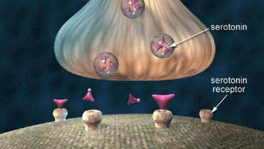
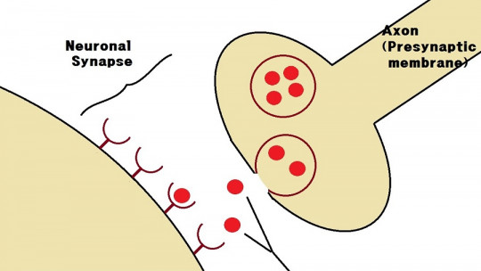
In the brain, receptors of this type are found on synaptic terminals regulating the release of neurotransmitters, both from its own receptors and those of other neurons.
Acetylcholine is a neurotransmitter that can be found in the brain, although it is also found in other parts of the body such as neuromuscular junctions and ganglia. In the case of muscarinic receptors, they fulfill the following functions.
1. Recovery receptors
Acetylcholine is always used as the neurotransmitter within the autonomic ganglion The nicotinic receptors of the postganglionic neuron are responsible for the rapid initial depolarization of the neuron.
After this process, there is a hyperpolarization of the neuron, followed by a slow depolarization, which represents a recovery period for the postganglionic neuron. This process is mediated by muscarinic receptors M1 and M2.
2. Postganglionic neurons
muscarinic receptors They are present at the junction of innervated tissues and postganglionic neurons of the parasympathetic system since acetylcholine is also found in this subsystem of the autonomic system.
3. Innervated tissue
Some parts of the sympathetic system use cholinergic receptors. This is the case of sweat glands, whose receptors are muscarinic type.
In the somatic nervous system, nicotinic receptors for acetylcholine are used at neuromuscular junctions.
Types of muscarinic receptors
Muscarinic receptors belong to the group of metabotropic receptors that use G proteins as a signaling mechanism. In these receptors, the molecule or ligand used to give the signal binds to the receptor, which has seven transmembrane regions. In the case of muscarinic receptors, the ligand is acetylcholine.
Up to five different types of muscarinic receptors have been discovered, which are named “M” followed by a number between 1 and 5. The M1, M3 and M5 receptors couple to Gq proteins, while M2 and M4 do so. made with Gi/o proteins.
Studying the chromosomes, geneticists and molecular biologists have discovered five genes that are involved in encoding muscarinic receptors , being named in the same way as the receivers but with the letter “m” in lower case. The genes m1, m2, m3 and m4 encode muscarinic M receptors 1 to 4. M5 is a type of receptor subtype that has not yet been detected pharmacologically.
1. M1 Receiver
This receptor is found mediating the slow excitatory postsynaptic potential (ESPS) of the ganglion in the postganglionic nerve. It is common in exocrine glands and the central nervous system. It is mostly bound to Gq-type proteins.
2. M2 Receiver
M2 receivers They are found in the heart, where they are responsible for slowing down the heartbeat, keeping it below the normal rate They do this by slowing down the rate of depolarization.
In humans, when we are resting, vagal activity dominates sympathetic activity. If M2 receptors are inhibited, then the heart rate increases.
3. M3 Receiver
The M3 receptor can be found in various places in the body. They are found in the muscles responsible for the contraction of blood capillaries and also in the lungs As with M1 receptors, M3 are Gq-type proteins.
4. M4 receiver
The M4 receptor is found mainly in the central nervous system and has inhibitory functions If stimulated with muscarinic agonists, bronchospasm may occur.
5. M5 receiver
The location of the M5 receivers is not completely known. As with the M1 and M3 receptors, M5 couples to Gq proteins.
Clinical importance
Different brain functions are known in which acetylcholine and its receptors, including muscarinic receptors, are involved. This can be observed in some pathologies, related to alterations in cholinergic transmission, being notable the case of neurodegenerative diseases such as Alzheimer’s or Parkinson’s disease.
In 1976, the first biochemical abnormality associated with Alzheimer’s disease was known. It was seen that in the hippocampus and cerebral cortex of the patients the enzyme choline acetyltransferase (CAT) was present at levels well below normal This enzyme catalyzes the synthesis of acetylcholine from its precursor substances: choline and acetylcoenzyme A.
Alzheimer disease
The fact that there is less activity in the CAT indicates that there is a loss of cholinergic nerve terminals that release acetylcholine in brain regions which, once they degenerate, are associated with the symptoms of Alzheimer’s. The regions of greatest deficit are the basal nucleus of Meynert and the temporal lobes.
In the case of this particular disease, the M2 receptor and nicotinic receptors, which are also sensitive to acetylcholine, are altered, while the M1, which is present in the hippocampus, is more or less preserved. Other neurotransmitters are also involved in Alzheimer’s disease, such as serotonin, glutamate, GABA, norepinephrine and somatostatin.
Biochemical abnormalities regarding acetylcholine in the hippocampus have been linked to the most well-known symptom of the disease: memory loss. The cholinergic terminals of the hippocampus are very important for memory formation and, therefore, The cognitive defects of the disease are related to problems in the function of muscarinic receptors in this region and the synthesis of the neurotransmitter.