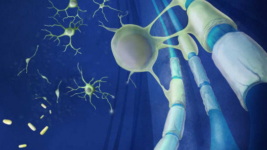
When we think about the cells of the human brain and the nervous system In general, the image of the neurons. However, these nerve cells by themselves cannot form a functional brain: they need the help of many other “pieces” with which our body is built.
The myelin, for example, is part of those materials without which our brain could not carry out its operations effectively. And neurons are supported by other components of the nervous system that fulfill discrete but at the same time important functions, as we will see in this article about myelin and its characteristics.
What is myelin?
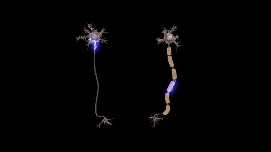
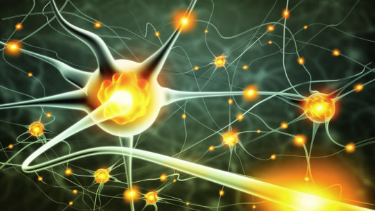
When graphically representing a neuron, either through a drawing or a 3D model, we normally draw the area of the nucleus, the branches with which it connects to other cells and an extension called the axon that is used to reach distant areas. However, in many cases that image would be incomplete. Many neurons have, around their axons, a whitish material that insulates them from the extracellular fluid. This substance is myelin, and its presence is essential for the proper functioning of the nervous system.
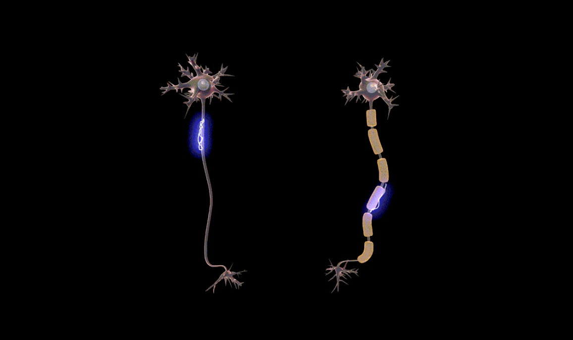
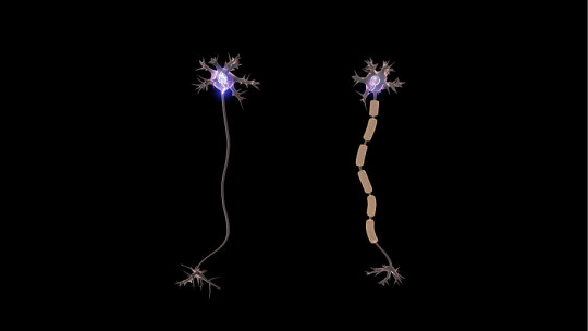
Myelin is a thick lipoprotein layer (made up of fatty substances and proteins) that surrounds the axons of some neurons, forming sausage or roll-shaped sheaths. These myelin sheaths have a very important function in our nervous system: allow the transmission of nerve impulses quickly and efficiently between the nerve cells of the brain and the spinal cord This material creates a kind of insulating layer around the projection of the neuron, through which it is important that electricity be transmitted quickly.
The function of myelin
The electrical current that passes through neurons is the type of signal with which these nerve cells function. Myelin allows these electrical signals to spread very quickly through the axons, so that this stimulus reaches the spaces in which neurons communicate with each other in time. In other words, the main added value that these sheaths provide to the neuron is the speed in the propagation of electrical signals.
If we removed an axon’s myelin sheaths, the electrical signals traveling along it would be much slower or could even be lost along the way. Myelin acts as an insulator, so that the current does not dissipate outside the path and only goes inside the neuron.
Ranvier’s nodes
The myelinated layer that covers the axon is called the myelin sheath, but it is not completely continuous along the axon, but between the myelinated segments there are uncovered regions. These areas of the axon that remain in contact with the extracellular fluid are called nodes of Ranvier .
The existence of the nodes of Ranvier is important, since without them the presence of myelin would be of no use. In these spaces, the electric current that spreads through the neuron gains strength, since in the nodes of Ranvier there are ion channels that, by acting as regulators of what enters and leaves the neuron, allow the signal not to be lost. force.
The action potential (nervous impulse) jumps from one node to another because these, unlike the rest of the neuron, are equipped with groups of sodium and potassium channels, so that the transmission of nervous impulses is more efficient. fast. The interaction between the myelin sheath and the nodes of Ranvier allows the nervous impulse to travel with greater speed, in a Saltatory manner (from one node of Ranvier to the next) and with less possibility of error.
Where is myelin found?
Myelin is present in the axons of many types of neurons, both in the Central Nervous System (that is, the brain and spinal cord) and outside of it. However, in some areas its concentration is higher than in others. Where myelin is abundant, it can be seen without the help of a microscope.
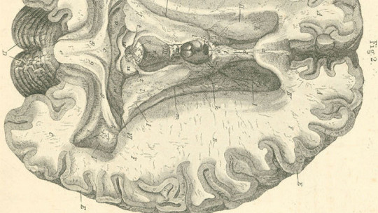
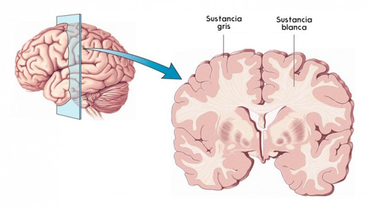
When we describe a brain it is common to talk about gray matter, but also, and although this fact is somewhat less known, there is the white matter The areas in which white matter is found are those in which myelinated neuronal bodies are so abundant that they change the color of those areas seen with the naked eye. That is why the areas in which the nuclei of neurons are concentrated usually have a grayish color, while the areas through which the axons essentially pass are white.
Two types of myelin sheaths
Myelin is essentially a material that serves one function, but there are different cells that form myelin sheaths. The neurons that belong to the Central Nervous System have myelin layers formed by a type of cells called oligodendrocytes, while the rest of the neurons use bodies called Schwann cells . Oligodendrocytes are shaped like a sausage crossed from end to end by a rope (the axon), while Scwann cells wrap the axons in a spiral, acquiring a cylindrical shape.
Although these cells are slightly different, they are both glial cells with a practically identical function: forming myelin sheaths.
Diseases due to myelin alteration
There are two types of diseases that are related to abnormalities in the myelin sheath: demyelinating diseases and dysmyelinating diseases
Demyelinating diseases are characterized by a pathological process directed against healthy myelin, unlike dysmyelinating diseases, in which there is inadequate formation of myelin or an impairment of the molecular mechanisms to maintain it in its normal conditions. The different pathologies of each type of disease related to myelin alteration are:
Demyelinating diseases
Dysmyelinating diseases
To learn more about myelin and its associated pathologies
Below we leave you an interesting video about Multiple Sclerosis, which explains how myelin is destroyed during the course of this pathology:


