

See more of our articles in your search results.
Think about the last time you solved a tricky problem at work, weighed a difficult decision, or simply followed a complicated conversation while planning your response at the same time. All of that mental effort depends heavily on the neocortex, the thin, folded outer layer of the brain responsible for most of what we consider distinctly human thought. It handles language, abstract reasoning, planning, and conscious perception, layering sophisticated cognitive capacity on top of the more automatic processes managed elsewhere in the brain. Most people never think about this structure directly, yet it shapes nearly every deliberate choice they make in a day. Understanding what it actually does, and how it differs from other brain regions, clarifies a great deal about human cognition.
The confusion many readers run into comes from how casually terms like “neocortex,” “cerebral cortex,” and “gray matter” get used interchangeably online, often without much precision. Some explanations oversimplify the neocortex into a single all-purpose “thinking center,” which glosses over just how specialized different regions actually are. Others focus so heavily on abstract evolutionary theory that the practical, everyday relevance of the structure gets lost entirely. This matters because the neocortex is not a vague metaphor; it is a real, densely layered structure whose specific regions handle distinct jobs, from processing vision to regulating impulse control.
What exactly makes this part of the brain so central to human cognition? The details are more specific than most casual explanations suggest.
This article covers what the neocortex is, its structural layers and major regions, the functions it supports, and how it develops and changes across a person’s lifespan.

How many of these have you been "meaning to read" for months?
- 1
 TyndaleThe Life-Changing Art of Self-Brain SurgeryConnecting Neuroscience and Faith to Radically Transform Your LifeListen Free On Audible
 - 2
 American Psychiatric AssociationDiagnostic and Statistical Manual of Mental Disorders, Text Revision Dsm-5-trGeneric jeufunListen Free On Audible
 - 3
 Ballantine BooksHollow BonesA NovelListen Free On Audible
What is the neocortex and where is it located?
The neocortex is the outermost layer of the mammalian brain, a thin sheet of tightly folded tissue that covers the cerebral hemispheres and handles higher-order functions like reasoning, language, and conscious perception. It sits above older, deeper brain structures.
Structurally, the neocortex forms the visible, wrinkled surface most people picture when they imagine a human brain. Those wrinkles, known as gyri and sulci, exist because the neocortex is far larger in surface area than the skull could otherwise accommodate, so it folds in on itself repeatedly to fit. This folding pattern increases the total amount of processing tissue considerably, which researchers generally connect to the expanded cognitive capacities seen in humans and other highly intelligent mammals.
The term “neo” reflects the structure’s relatively recent evolutionary appearance compared to older brain regions like the brainstem and parts of the limbic system. Neuroscientist Paul MacLean, who proposed the historically influential triune brain theory, described the neocortex as the newest of three evolutionary brain layers, sitting above a limbic layer and a deeper brainstem layer. While that specific three-layer model is now considered outdated in several respects, the basic observation that the neocortex expanded dramatically in more recent evolutionary history remains broadly accurate.
Comparative brain size gives a sense of just how significant this structure is. In humans, the neocortex makes up a very large proportion of total brain volume, far more than in most other mammals, which helps explain why so much cognitive research focuses specifically on this single structure rather than the brain as a whole.

What are the main structural layers of the neocortex?
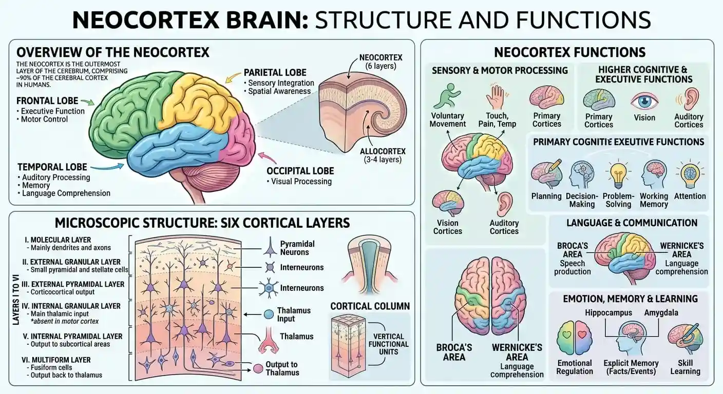
The neocortex is organized into six distinct horizontal layers, each containing different types of neurons and serving a different role in receiving, processing, and transmitting information. This layered architecture is a defining structural feature of the region.
Layer one, closest to the brain’s surface, contains relatively few neuron cell bodies and mainly consists of connecting fibers. Layers two and three handle much of the communication between different cortical regions, essentially acting as relay points for information moving across the brain’s surface. Layer four typically receives incoming sensory information from deeper brain structures like the thalamus, making it especially prominent in sensory processing regions such as the visual cortex. Layers five and six send output signals to other brain regions and to structures outside the cortex entirely, including the spinal cord in some cases.
This six-layer structure is remarkably consistent across most of the neocortex, though the relative thickness of each layer varies depending on the region’s specific function. Sensory areas tend to have a thicker layer four, since they are built to receive dense incoming signals, while motor areas often have a thicker layer five, reflecting their role in sending commands outward. Small structural differences like these tell researchers a great deal about what a given region actually does.
| Cortical layer | Primary role |
|---|---|
| Layer I | Contains mostly connecting fibers with few neuron cell bodies. |
| Layers II-III | Support communication between different cortical regions. |
| Layer IV | Receives incoming sensory signals, especially from the thalamus. |
| Layers V-VI | Send output signals to other brain regions and beyond the cortex. |
Understanding this layered organization helps explain why damage to even a small area of the neocortex can produce very specific, localized deficits rather than a generalized loss of function.
What are the four lobes of the neocortex and their functions?
The neocortex is divided into four major lobes, the frontal, parietal, temporal, and occipital lobes, each associated with a distinct set of cognitive and sensory functions. This division provides a practical map for understanding cortical organization.
The frontal lobe sits at the front of the brain and handles planning, decision-making, impulse control, and much of what psychologists call executive function. Neuroscientist and psychologist Antonio Damasio has spent decades studying how damage to specific frontal regions, particularly the prefrontal cortex, can dramatically alter personality and decision-making even when basic intelligence remains intact. His research helped demonstrate that emotion and rational judgment are far more intertwined within this lobe than earlier theories assumed.
The parietal lobe, located toward the top and back of the brain, integrates sensory information from touch, spatial awareness, and body position, helping construct a coherent sense of where your body is relative to the world around it. The temporal lobe, positioned near the ears, plays a central role in auditory processing, language comprehension, and aspects of long-term memory formation, working closely with deeper structures like the hippocampus. The occipital lobe, at the very back of the brain, is dedicated almost entirely to visual processing, transforming raw signals from the eyes into recognizable shapes, colors, and movement.
- Frontal lobe functions include planning, impulse control, and complex decision-making.
- Parietal lobe functions include spatial awareness and integration of sensory information.
- Temporal lobe functions include language processing and aspects of memory formation.
- Occipital lobe functions center almost entirely on visual processing and interpretation.
What functions does the neocortex control?
The neocortex controls most higher-order cognitive functions, including language, abstract reasoning, conscious perception, voluntary movement, and complex planning. These capacities generally require far more processing than the automatic functions handled by deeper brain structures.
Language offers a clear illustration of this specialization. Specific neocortical regions, generally located in the left hemisphere for most right-handed individuals, handle distinct aspects of language production and comprehension. Damage to one region might leave a person able to understand speech perfectly while struggling to produce fluent sentences, while damage to a nearby but distinct region can produce the opposite pattern entirely. This double dissociation has taught researchers an enormous amount about how finely specialized different neocortical areas really are.
Neuroscientist Nancy Kanwisher, known for research on functional specialization within the visual cortex, has shown that certain small cortical regions respond selectively to very specific categories of visual information, such as faces, rather than processing all visual input identically. This kind of fine-grained specialization runs throughout the neocortex, not just in visual regions, and it helps explain why even subtle brain injuries can produce surprisingly specific cognitive changes rather than a uniform decline across all mental abilities.
- Sensory input is received by specialized regions like the visual or auditory cortex.
- Information is integrated across association areas that combine sensory and contextual data.
- Reasoning and planning occur primarily within frontal regions of the neocortex.
- Voluntary action is initiated through motor cortex regions that send commands to the body.

How does the neocortex differ from the limbic system and brainstem?
The neocortex differs from the limbic system and brainstem mainly in the type of processing it performs, handling complex, conscious, and often deliberate cognition rather than the automatic emotional and physiological regulation managed by deeper brain structures. All three regions work together continuously.
The brainstem, often associated informally with the term “reptilian brain,” manages automatic survival functions like heart rate, breathing, and basic reflexes, operating largely outside conscious control. The limbic system, including structures like the amygdala and hippocampus, adds emotional weight and memory context to incoming information, shaping how a situation feels rather than how it is logically analyzed. The neocortex builds on both of these layers, adding language, abstract planning, and the kind of deliberate, effortful reasoning that lets a person override an immediate emotional impulse when the situation calls for it.
This does not mean the neocortex simply overrides emotion in some clean, hierarchical way. Damasio’s research specifically challenged that assumption, showing that people with damage to certain frontal neocortical regions often struggle with decision-making precisely because they lose access to emotional signals that normally guide sound judgment. Purely “rational” thought, disconnected entirely from emotion, often turns out to function poorly rather than optimally.
A useful way to think about this relationship is as three deeply interconnected systems rather than three separate brains stacked on top of each other. Fast. Slow. Integrated. Signals constantly move between these regions, blending automatic reflex, emotional context, and deliberate reasoning into a single, continuous response rather than three isolated processes running independently.
How do the two brain hemispheres divide neocortical functions?
The neocortex is split into two hemispheres that share many functions but also show some meaningful specialization, particularly around language and certain types of spatial or holistic processing. This division does not follow the popular “left brain, right brain” personality stereotype.
Neuroscientist Michael Gazzaniga, whose pioneering research on patients whose hemispheres had been surgically separated shaped much of modern understanding of hemispheric specialization, demonstrated that each hemisphere can process certain types of information somewhat independently under specific experimental conditions. His work showed that language functions are typically concentrated in the left hemisphere for most people, while certain aspects of spatial reasoning and facial recognition tend to rely more heavily on the right hemisphere. Crucially, Gazzaniga’s research also emphasized that the two hemispheres constantly communicate through a thick band of fibers called the corpus callosum during normal, everyday functioning.
The popular idea that people are predominantly “left-brained” or “right-brained,” with one hemisphere driving their entire personality or thinking style, does not hold up under scientific scrutiny. Most complex tasks, including language and creative problem-solving, draw on both hemispheres working together rather than one dominating exclusively. This is an area where oversimplified pop psychology has significantly outpaced the actual neuroscience.
A practical takeaway here is straightforward: hemispheric specialization is real and well documented for specific, narrow functions, but it does not translate into broad personality categories. Treating “left-brained” or “right-brained” as a meaningful description of someone’s overall thinking style misrepresents decades of careful hemisphere research.
How does the neocortex support memory, language, and reasoning?
The neocortex supports memory, language, and reasoning by coordinating with deeper structures like the hippocampus while performing the more complex integration, storage, and retrieval processes associated with long-term and abstract thinking. These functions rely on extensive networks rather than single, isolated regions.
Long-term memory formation depends on continuous interaction between the hippocampus and specific neocortical regions, particularly during sleep, when memories are thought to be gradually transferred and consolidated into more stable cortical storage. This is one reason chronic sleep disruption can noticeably affect memory retention over time; the neocortex simply has less opportunity to complete this consolidation process. Reasoning and abstract thought rely heavily on the prefrontal cortex, which helps weigh competing options, delay gratification, and hold multiple pieces of information in mind simultaneously.
Neuroscientist Michael Posner, known for research on attention networks in the brain, has shown that sustained, focused attention depends on specific neocortical and subcortical circuits working in coordination, rather than a single generalized “attention center.” This research has practical implications for anyone struggling with focus or distraction, since it suggests that attention is a trainable, network-based skill rather than a fixed personal trait.
- Memory consolidation relies on ongoing communication between the hippocampus and neocortex, especially during sleep.
- Working memory depends heavily on prefrontal cortex activity during complex problem-solving.
- Language processing draws on specialized temporal and frontal regions working in close coordination.
- Sustained attention relies on distributed neural networks rather than one isolated brain region.
What happens when the neocortex is damaged or underdeveloped?
Damage to the neocortex can produce highly specific cognitive or behavioral changes depending on which region is affected, ranging from language difficulties to impaired decision-making or altered personality, rather than a single uniform decline across all abilities.
Neuroscientist V.S. Ramachandran, well known for his research on sensory remapping and conditions like phantom limb sensation, has documented cases where damage to specific cortical regions produces strikingly unusual and highly localized symptoms. His work illustrates just how precisely mapped the neocortex actually is, since even relatively small areas of damage can produce very particular deficits, such as difficulty recognizing faces while other visual abilities remain fully intact.
Frontal lobe damage, in particular, has been studied extensively because of its effects on personality, impulse control, and social judgment. People with significant frontal damage sometimes retain normal intelligence test scores while showing dramatic changes in planning ability, emotional regulation, or social behavior, a pattern that has taught researchers a great deal about how distinct cognitive functions really are from one another. This distinction matters clinically, since it means standard intelligence measures alone cannot capture every important aspect of someone’s cognitive functioning after a brain injury.
It is worth being clear that recovery and adaptation after neocortical damage vary enormously between individuals, depending on factors like age, injury location, and rehabilitation support. Anyone experiencing changes in thinking, memory, or behavior following an injury or illness should consult a qualified medical or neuropsychological professional for individualized assessment rather than relying on general information like this article to interpret their specific situation.
How does the neocortex develop across the lifespan?
The neocortex develops gradually from before birth through early adulthood, with different regions maturing at different rates, and it continues to change structurally in smaller ways throughout later adulthood. Frontal regions generally mature last.
Early neocortical development begins during prenatal growth, when neurons form and begin migrating to their eventual cortical locations, establishing the basic six-layer structure well before birth. Sensory and motor regions tend to mature relatively early during childhood, which is part of why basic movement and perceptual skills develop faster than more complex reasoning abilities. The prefrontal cortex, responsible for planning, impulse control, and long-term decision-making, continues developing well into a person’s twenties, a finding with real implications for how adolescent risk-taking and decision-making are understood.
This extended developmental timeline helps explain certain well-documented patterns in adolescent behavior, including a tendency toward impulsivity and heightened sensitivity to social reward, since the brain regions responsible for regulating those impulses are simply still under construction during the teenage years. It does not mean adolescents lack reasoning ability altogether; it means the balance between impulse and regulation is still shifting during this developmental period.
In later adulthood, the neocortex undergoes gradual structural changes, including modest thinning in certain regions, though the pace and pattern of this change vary considerably between individuals based on genetics, health, and lifestyle factors. Healthy aging does not automatically mean significant cognitive decline, and ongoing engagement with learning, social interaction, and physical activity appears associated with better maintained cognitive function later in life, according to a substantial body of aging research.
How can you support healthy neocortex function?
You can support healthy neocortex function through consistent sleep, regular physical activity, ongoing mental engagement, and social connection, all of which are associated with better cognitive performance and structural brain health across large population studies. No single habit guarantees a specific outcome.
Sleep deserves particular attention here, since it plays a direct role in memory consolidation processes that depend heavily on coordinated activity between the neocortex and hippocampus. Chronic sleep deprivation has been linked repeatedly to impaired attention, slower reasoning, and difficulty forming new long-term memories, effects that tend to improve once healthier sleep patterns are restored. Physical exercise also shows a strong, consistent association with cognitive health, likely through improved blood flow and the release of factors that support neural growth and connectivity throughout the cortex.
Mental engagement matters as well, though the specific claim that any single “brain training” app produces broad, lasting cognitive improvement remains scientifically contested. What does appear well supported is that genuinely novel, effortful learning, such as picking up an unfamiliar skill or language, tends to engage cortical networks more thoroughly than passive or highly repetitive activities. Social connection rounds out this picture, since maintaining meaningful relationships appears linked to slower cognitive decline in numerous long-term studies, possibly because social interaction demands flexible, complex cognitive processing on an ongoing basis.
None of these strategies function as guaranteed protection against cognitive decline or neurological illness, and genuine concerns about memory, attention, or thinking changes deserve evaluation by a qualified healthcare professional rather than self-directed lifestyle changes alone. Reasonable habits, applied consistently, still represent one of the most evidence-based ways to support long-term cognitive health available to most people.
FAQs about the neocortex
What is the main function of the neocortex?
The main function of the neocortex is to support higher-order cognitive processes, including language, abstract reasoning, planning, conscious perception, and voluntary movement control. Unlike deeper brain structures that manage automatic survival functions or basic emotional processing, the neocortex is specialized for the kind of complex, often deliberate thinking that humans rely on for problem-solving, communication, and long-term planning. Different regions within the neocortex handle distinct tasks, so damage or dysfunction in one area does not necessarily affect other cognitive abilities. For example, a person could experience difficulty with language processing after localized damage while retaining normal visual perception, memory, or motor control. This specialization is one of the defining features that separates the neocortex from more generalized, automatic brain regions like the brainstem.
Is the neocortex the same as the cerebral cortex?
The terms overlap considerably but are not perfectly interchangeable. The cerebral cortex refers broadly to the outer layer of the cerebral hemispheres, and in humans, the vast majority of that cortex is neocortex, meaning it has the characteristic six-layer structure. However, small portions of the cerebral cortex, such as parts of the hippocampus and some evolutionarily older cortical regions, are technically classified separately as allocortex because they have a different, simpler layered structure. In everyday usage, and in most general psychology writing, people often use “cerebral cortex” and “neocortex” somewhat interchangeably since the neocortex makes up such a dominant portion of the total cortical surface. Strictly speaking, though, neocortex refers specifically to the six-layered variety of cortical tissue.
How is the neocortex different from the reptilian brain?
The neocortex and the structures popularly labeled the “reptilian brain,” mainly the brainstem, differ in both evolutionary age and function. The brainstem manages automatic, largely unconscious survival processes like heartbeat, breathing, and basic reflexes, operating with minimal conscious involvement. The neocortex, by contrast, supports language, abstract reasoning, and deliberate decision-making, processes that require conscious effort and complex neural integration. These two regions are not isolated from each other; they constantly exchange signals, with the neocortex often adding context, memory, and reasoning on top of more automatic responses initiated by deeper brain structures. It is worth noting that the popular “reptilian brain” framing itself is considered an oversimplified model by many contemporary neuroscientists, even though the underlying brainstem anatomy it refers to is entirely real.
Can the neocortex repair itself after injury?
The neocortex has some capacity for reorganization and adaptation after injury, a property researchers call neuroplasticity, though the degree of recovery varies significantly depending on the injury’s location, size, and the person’s age and overall health. In some cases, nearby or connected regions can partially compensate for lost function through rewiring existing neural connections, particularly with the support of targeted rehabilitation like speech or occupational therapy. Recovery is rarely complete or guaranteed, however, and outcomes differ enormously from person to person. Younger individuals generally show somewhat greater plasticity than older adults, though meaningful improvement remains possible across a wide age range with appropriate treatment. Anyone recovering from a brain injury should work directly with qualified medical and rehabilitation professionals, since recovery plans need to be tailored to the specific injury and individual involved.
Why do humans have a larger neocortex than other animals?
Humans have a proportionally larger neocortex relative to overall brain size than most other animals, a difference researchers generally connect to the expanded cognitive capacities associated with complex language, abstract reasoning, and advanced social behavior. Some primates and other highly social mammals also show relatively large neocortical proportions, which has led some researchers to propose connections between neocortex size and the cognitive demands of navigating complex social relationships. It is worth noting that raw neocortex size alone does not fully explain intelligence differences between species, since factors like neural density, connectivity patterns, and specific regional development also matter considerably. The relationship between brain structure and cognitive capacity remains an active and evolving area of neuroscience research rather than a fully settled question.
Does the neocortex control emotions?
The neocortex plays a significant supporting role in emotional experience, but it does not control emotion in isolation. Core emotional processing relies heavily on subcortical structures like the amygdala, while the neocortex, particularly the prefrontal cortex, contributes context, interpretation, and regulation to those emotional signals. This is why a person might feel an initial surge of anger almost instantly, generated largely by faster subcortical processing, before slower neocortical regions help evaluate the situation and shape a more measured response. Research has shown that emotion and rational thought are far more intertwined within the brain than older models suggested, with damage to certain frontal neocortical regions sometimes impairing both emotional regulation and decision-making simultaneously. Emotion and cognition function as an integrated system rather than two separate, competing brain processes.
What happens to the neocortex as people age?
As people age, the neocortex generally undergoes some gradual structural changes, including modest thinning in certain regions and changes in connectivity between different areas. The pace and impact of these changes vary considerably from person to person, influenced by genetics, cardiovascular health, activity levels, and other lifestyle factors. Some cognitive functions, particularly those relying heavily on processing speed, may show mild decline for many people during normal aging, while other abilities, such as accumulated knowledge and certain types of reasoning, often remain stable or even improve well into later adulthood. Significant, rapid, or highly disruptive cognitive changes are not considered a normal or inevitable part of aging and warrant evaluation by a healthcare professional, since they may reflect a treatable underlying condition rather than typical age-related change.
How does the neocortex relate to intelligence?
The relationship between the neocortex and intelligence is genuine but more complex than simply “bigger means smarter.” Research suggests that factors like the efficiency of neural connections, the density of certain cortical regions, and the strength of communication between different cortical areas correlate more consistently with measured intelligence than raw cortical size alone. Different cognitive abilities, such as verbal reasoning, spatial processing, and working memory, also appear to rely on somewhat distinct neocortical networks rather than a single unified “intelligence center.” This helps explain why someone might show exceptional ability in one cognitive domain while performing more average in another, since intelligence is not a single uniform property generated by one part of the brain. Ongoing research continues to refine understanding of exactly how structural and functional cortical differences relate to the varied abilities that intelligence testing attempts to capture.
Bibliography
- MacLean, P. D. (1990). The Triune Brain in Evolution: Role in Paleocerebral Functions. Springer.
- Damasio, A. (1994). Descartes’ Error: Emotion, Reason, and the Human Brain. Putnam.
- Gazzaniga, M. S. (2015). Tales from Both Sides of the Brain: A Life in Neuroscience. Ecco.
- Ramachandran, V. S., & Blakeslee, S. (1998). Phantoms in the Brain: Probing the Mysteries of the Human Mind. William Morrow.
- Kanwisher, N. (2000). Domain specificity in face perception. Nature Neuroscience.
- Posner, M. I., & Petersen, S. E. (1990). The attention system of the human brain. Annual Review of Neuroscience.
- National Institute of Mental Health. The teen brain: Seven things to know. NIMH.
- American Psychological Association. Brain development and cognitive aging resources. APA.
- Mayo Clinic. Brain anatomy and function overview. Mayo Clinic.
Use this citation format to reference the article clearly and help readers find the original source.
PsychologyFor. (2026). Neocortex Brain: Structure and Functions. PsychologyFor. https://psychologyfor.com/neocortex-brain-structure-and-functions/