

See more of our articles in your search results.
Imagine losing the ability to feel your own leg move, or suddenly struggling to control your bladder after a head injury, with no obvious explanation from the tests your doctor initially runs. It’s a frightening, disorienting experience, made worse by how rarely anyone talks about the specific brain structure responsible for it. Most people have heard of the frontal lobe or the parietal lobe, but almost no one has heard of the small, U-shaped strip of cortex tucked between them that quietly governs leg movement, lower-body sensation, and even bladder control: the paracentral lobe.
This obscurity can leave patients and caregivers feeling lost after a stroke, tumor, or injury affects this exact region, since symptoms like leg weakness paired with incontinence don’t fit the more familiar narratives of memory loss or speech difficulty typically associated with brain damage. The confusion is compounded by the fact that this structure sits deep on the medial surface of the brain, folded into the crease between the two hemispheres, making it far less intuitive to visualize than the lobes on the brain’s outer surface. Understanding exactly where this structure lives and what it does can transform bewildering, seemingly disconnected symptoms into a coherent clinical picture. It also validates an experience that is often minimized simply because it’s unfamiliar.
Why does one small strip of cortex control something as basic as walking and something as personal as continence at the same time?
This article breaks down the anatomy, characteristics, and everyday functions of the paracentral lobe, along with what happens when this specific brain region is damaged.
What Is the Paracentral Lobe and Where Is It Located?
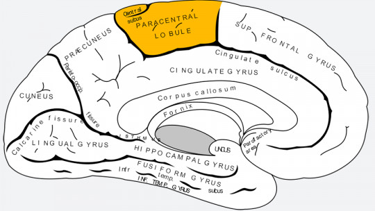
The paracentral lobe, more precisely called the paracentral lobule, is a U-shaped fold of cerebral cortex located on the medial surface of each hemisphere, where the brain’s two sides face each other across the longitudinal fissure. It is not one of the four classic lobes taught in introductory anatomy; rather, it represents the medial continuation of two familiar structures — the precentral gyrus and the postcentral gyrus — as they wrap around the top edge of the brain and fold inward.
How many of these have you been "meaning to read" for months?
- 1
 TarcherThe Artist's Way30th Anniversary EditionListen Free On Audible
 - 2
 Atria BooksThe Courage to Be DislikedThe Japanese Phenomenon That Shows You How to Change Your Life and Achieve Real HappinessListen Free On Audible
 - 3
 Hay HouseBecoming SupernaturalHow Common People Are Doing the UncommonListen Free On Audible
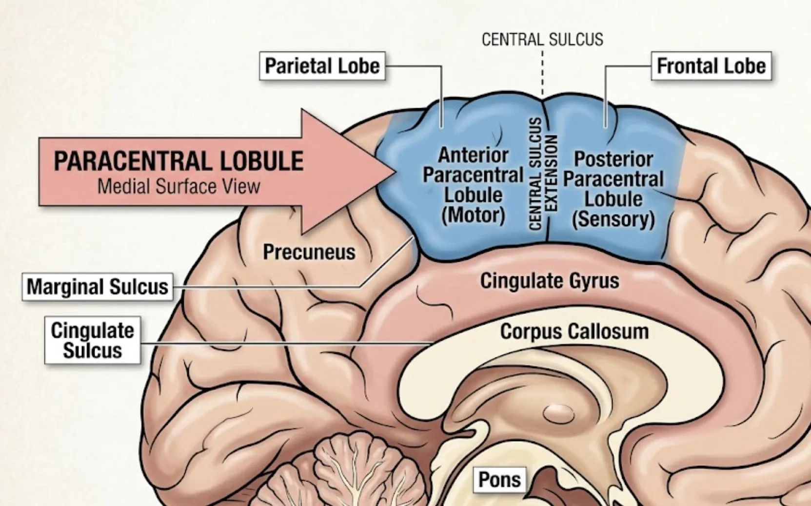
Because it straddles this fold, the paracentral lobule technically belongs to two different lobes at once: its anterior portion is part of the frontal lobe, continuing the primary motor cortex, while its posterior portion belongs to the parietal lobe, continuing the primary somatosensory cortex. The dividing line between these two halves is the continuation of the central sulcus (the sulcus of Rolando) onto the medial brain surface. Its anterior border is marked by the paracentral sulcus, while its posterior and inferior borders are formed by the cingulate sulcus and its marginal branch, which separate it from the precuneus behind.
Blood supply to this region comes from the anterior cerebral artery, a detail that carries real clinical weight, since strokes affecting this specific vessel produce a very recognizable and often misunderstood pattern of leg weakness disproportionate to arm or facial involvement, unlike the more commonly recognized middle cerebral artery stroke pattern.
Despite occupying a small surface area compared to the major lobes, the paracentral lobule punches well above its size in clinical importance, precisely because it represents such a concentrated, specific slice of the body’s motor and sensory map.
How Is the Paracentral Lobule Structured Anatomically?
The paracentral lobule is best understood as two adjoining halves working as a single functional unit. Its anterior half is the medial extension of the precentral gyrus, and its posterior half is the medial extension of the postcentral gyrus, joined seamlessly across the medial surface.
The anterior paracentral lobule contains the medial-most portion of the primary motor cortex, corresponding to Brodmann area 4, and blends into the supplementary motor area just in front of it, a region involved in planning and sequencing complex, self-initiated movements. The posterior paracentral lobule contains the medial-most portion of the primary somatosensory cortex, corresponding to Brodmann areas 3, 1, and 2, mirroring the same cytoarchitecture found on the lateral surface of the brain but dedicated to a different, more specific part of the body map.
- The anterior paracentral lobule extends the primary motor cortex onto the medial brain surface, controlling voluntary leg movement.
- The posterior paracentral lobule extends the primary somatosensory cortex, processing touch and proprioception from the lower limb.
- The paracentral sulcus marks its front boundary, separating it from the medial frontal gyrus.
- The marginal sulcus, an upward branch of the cingulate sulcus, marks its rear boundary, separating it from the precuneus.
This layout mirrors the broader organization first mapped by neurosurgeon Wilder Penfield, whose pioneering intraoperative stimulation studies charted how different body parts are represented across the motor and sensory strips, producing the now-famous cortical homunculus, a distorted human figure whose proportions reflect representational density rather than actual body size. The paracentral lobule occupies the very tip of that map, where the homunculus’s leg, foot, and genital regions are represented.
What Is the Motor and Sensory Homunculus in This Region?
The homunculus is a visual and conceptual map showing which body parts are controlled by which parts of the motor and sensory cortex, and it directly explains the paracentral lobule’s specialized role. Rather than an even distribution, cortical representation is proportional to how finely controlled or sensitive a body part needs to be, not its actual physical size.
Because the leg, foot, and perineal region require this medial extension of cortex to be represented, the paracentral lobule effectively houses the “feet and lower body” section of the classic homunculus figure. This is why the lower limb representation sits folded onto the medial surface rather than staying on the lateral convexity alongside the hand, face, and trunk. Neurophysiologist Vernon Mountcastle’s foundational research on the columnar organization of the somatosensory cortex helped establish that neurons throughout this region, including the paracentral lobule, are arranged in vertical functional columns, each processing a specific type of sensory input, such as light touch or joint position, for a highly localized part of the body.
This tight, columnar organization explains why damage to a very small area of the paracentral lobule can produce a symptom as narrow as numbness confined strictly to one foot or difficulty moving just the toes, rather than a broader, diffuse deficit. Clinicians use this predictable mapping diagnostically, since a patient’s exact pattern of leg weakness or sensory loss can help localize a lesion to the paracentral region even before imaging confirms it.
How Does the Paracentral Lobe Control Movement of the Legs?
The paracentral lobule is the single most important cortical region for voluntary movement of the lower limb. Its anterior half sends direct motor commands down through the corticospinal tract to control muscles in the thigh, lower leg, and foot on the opposite side of the body.
These descending motor signals travel through the internal capsule and brainstem before crossing to the opposite side at the medullary pyramids, which is why damage to one paracentral lobule produces weakness in the contralateral leg rather than the same-sided one — a crossover pattern shared with motor control throughout the rest of the primary motor cortex. The supplementary motor area adjoining the paracentral lobule contributes an additional layer, helping plan and sequence more complex movements like initiating a step or coordinating bilateral leg movements needed for tasks like climbing stairs.
Researcher Marc Jeannerod’s extensive work on motor cognition demonstrated that movement planning involves an internal, predictive model of the body constructed before a muscle ever contracts, a process that depends on close cooperation between motor regions like the paracentral lobule and the sensory feedback arriving from the body itself. This predictive quality is part of why damage to this region doesn’t just cause weakness; it can also disrupt the smoothness and timing of leg movements, producing an awkward, effortful gait even when some strength remains.
A useful practical marker for clinicians and physical therapists: leg weakness that is proportionally worse than arm weakness after a stroke or brain injury should immediately raise suspicion for a paracentral or anterior cerebral artery territory lesion rather than the more commonly recognized middle cerebral artery pattern.

How Does This Region Process Sensation From the Lower Body?
The posterior paracentral lobule is the primary cortical destination for touch, pressure, temperature, pain, and proprioceptive signals arising from the leg, foot, and lower body. Without it, this specific region of the body effectively goes “silent” to conscious perception, even though the peripheral nerves themselves remain perfectly intact.
Sensory information travels from receptors in the skin, muscles, and joints through the spinal cord and thalamus before arriving at the postcentral gyrus’s medial extension inside the paracentral lobule, where it is consciously registered and localized. Proprioception — the sense of where a limb is in space without looking at it — is especially dependent on this region, which is why damage here can leave a person unable to sense their leg’s position, causing them to misjudge steps or lose balance even when muscle strength is otherwise preserved.
Physician and neuroscientist Antonio Damasio’s broader research on body awareness and interoception highlighted how crucial an accurate, continuously updated internal body map is for a stable sense of self and effective movement, a principle that applies directly to how the paracentral lobule contributes to whole-body awareness. When this sensory feedback loop breaks down, people sometimes describe their own leg as feeling foreign, disconnected, or “not quite theirs,” a genuinely disorienting experience rather than an exaggeration.
Occupational and physical therapists addressing paracentral lobule damage often incorporate sensory re-education exercises, deliberately practicing tasks like identifying textures or joint positions without visual cues, to help retrain whatever residual capacity remains in this circuit.
What Is the Paracentral Lobule’s Role in Bladder and Bowel Control?
Perhaps the most surprising function of the paracentral lobule is its involvement in voluntary control of urination and defecation. This cortical region provides the conscious “permission” signal that allows a person to delay these reflexes until an appropriate time and place.
The pontine micturition center in the brainstem manages the basic reflex circuitry of bladder emptying, but the paracentral lobule exerts top-down voluntary control over that reflex, allowing humans to override the urge to urinate or defecate until socially and practically appropriate. This is a distinctly cortical, learned capacity rather than a purely automatic one, which is part of why toddlers develop continence gradually as this cortical control matures, and why certain neurological injuries can cause it to regress.
Bilateral damage to the paracentral lobules, most classically seen with strokes affecting both anterior cerebral arteries or with tumors compressing this medial region, can produce urinary incontinence alongside bilateral leg weakness, a combination that should immediately prompt clinicians to consider this specific location rather than a more generalized or peripheral cause. This connection is frequently under-recognized outside of neurology, leaving patients and families confused when incontinence appears alongside leg symptoms rather than being attributed simply to “getting older” or an unrelated urological problem.
- Voluntary bladder control depends on cortical override signals originating largely from the paracentral lobule.
- Bilateral paracentral damage can produce combined leg weakness and incontinence, a distinctive diagnostic pattern.
- Continence development in early childhood reflects the gradual maturation of this same cortical control system.

How Does the Paracentral Lobule Connect to the Supplementary Motor Area?
The paracentral lobule does not work alone; it sits immediately adjacent to the supplementary motor area, and the two regions cooperate closely to plan and execute coordinated, self-generated movement. This partnership becomes especially important for movements that require timing and internal initiation rather than a simple external trigger.
The supplementary motor area is particularly active in preparing sequences of movement before they occur, such as planning the multiple steps needed to walk across a room or coordinating both legs during more complex activities like climbing stairs. Neuroscientist Giacomo Rizzolatti’s broader body of research on the motor system, including work on how the brain represents and organizes goal-directed action, helped establish that regions like the supplementary motor area function as part of an integrated network for movement planning, rather than operating as isolated modules disconnected from surrounding cortex like the paracentral lobule.
This close anatomical and functional relationship explains a clinically important pattern: damage limited to the supplementary motor area alone can cause a temporary difficulty initiating movement, sometimes called supplementary motor area syndrome, that often improves substantially over weeks, whereas damage to the paracentral lobule itself tends to produce more persistent weakness, since it houses the actual primary motor output pathway rather than just the planning stage.
Distinguishing between these two nearby but functionally distinct regions matters enormously for prognosis, since a family told simply “there’s damage near the top of the brain” may reasonably fear permanent paralysis, when in fact the specific structure involved carries a meaningfully different recovery trajectory.
What Happens When the Paracentral Lobule Is Damaged?
Damage to the paracentral lobule most commonly produces a distinctive triad: contralateral leg weakness, lower-limb sensory loss, and impaired voluntary bladder or bowel control. Recognizing this specific combination helps clinicians localize the injury even before advanced imaging is available.
The leading cause is stroke affecting the anterior cerebral artery, since this vessel supplies the medial brain surface where the paracentral lobule sits; falls, tumors compressing the medial hemisphere, and parasagittal meningiomas — tumors arising near the midline — are other recognized causes. Because the artery supplying this region can affect both hemispheres in some anatomical variants, bilateral symptoms are more common here than with strokes affecting the lateral cortex, which is part of why paracentral lobule injuries can look deceptively different from the classic one-sided stroke presentation most people picture.
- Contralateral leg weakness disproportionate to arm involvement is a hallmark sign of paracentral lobule damage.
- Lower-limb sensory loss, including impaired proprioception, often accompanies the motor deficit.
- Urinary incontinence can occur with bilateral damage, distinguishing this pattern from more lateral strokes.
- Gait disturbance frequently results from the combination of weakness and disrupted proprioceptive feedback.
Because this symptom pattern is less familiar to the general public than the arm weakness and facial drooping associated with more common strokes, patients and bystanders sometimes delay seeking emergency care, mistakenly attributing sudden leg weakness to a musculoskeletal problem rather than recognizing it as a possible neurological emergency.

How Are Paracentral Lobule Conditions Diagnosed and Treated?
Diagnosis relies on recognizing the specific pattern of leg-predominant weakness combined with sensory changes and, in bilateral cases, bladder involvement, followed by targeted imaging to confirm the location and cause. Because the symptom pattern is distinctive, an experienced clinician can often localize the problem to this region during a bedside neurological examination alone.
MRI is the preferred imaging tool for visualizing the medial brain surface and identifying strokes, tumors, or other structural lesions affecting the paracentral region, since this area can be more difficult to appreciate on standard CT imaging due to its location deep within the interhemispheric fissure. When a stroke is suspected, urgent vascular imaging of the anterior cerebral artery helps confirm the diagnosis and guides acute treatment decisions, including the potential use of clot-dissolving medication within the appropriate treatment window.
Rehabilitation typically combines physical therapy focused on gait training and lower-limb strengthening with occupational therapy addressing sensory re-education and, when needed, structured bladder retraining programs. Recovery outcomes vary considerably depending on the size and cause of the lesion, but the brain’s capacity for functional reorganization means meaningful improvement is achievable for many patients, particularly with early, consistent rehabilitation engagement.
- Seek urgent evaluation for any sudden leg weakness, even without arm or facial symptoms, since this pattern can still indicate a stroke.
- Request targeted imaging of the medial brain surface and anterior cerebral artery when this specific symptom pattern is present.
- Begin gait and sensory rehabilitation promptly, since early, structured therapy improves functional outcomes.
- Address bladder symptoms openly with a clinician, since incontinence following brain injury is a recognized medical issue rather than a personal failing.
How Does the Paracentral Lobule Differ Across Development and Aging?
The paracentral lobule’s functions mature gradually in early childhood and can show measurable vulnerability in older adulthood, particularly related to vascular health. Understanding this trajectory helps set realistic expectations for both developmental milestones and age-related risk.
In early childhood, the gradual myelination of the corticospinal pathways originating in this region underlies the developmental progression from reflexive lower-limb movement in infancy toward coordinated walking and, eventually, voluntary bladder and bowel control, typically achieved between two and four years of age. This maturation timeline explains why continence training works best when approached with patience rather than pressure, since the underlying cortical control system genuinely needs time to develop rather than being simply a matter of willpower.
In older adults, cardiovascular risk factors such as hypertension, diabetes, and atherosclerosis increase vulnerability to strokes affecting the anterior cerebral artery territory, making the paracentral lobule a relevant consideration in vascular risk-reduction conversations, even though it rarely receives the same public attention as the arteries supplying more well-known brain regions. Age-related changes in proprioceptive processing throughout the sensory cortex, including the posterior paracentral lobule, also contribute to the increased fall risk seen in many older adults, independent of muscle strength alone.
Practical prevention strategies include managing blood pressure and blood sugar consistently, staying physically active to preserve gait and balance, and seeking prompt medical attention for any sudden change in leg strength, sensation, or bladder control rather than assuming it reflects ordinary aging.
FAQs about the Paracentral Lobe
What is the main function of the paracentral lobe?
The paracentral lobule’s primary functions are controlling voluntary movement of the leg and foot and processing sensation from the lower limb, including touch, temperature, and proprioception. It also plays an essential role in voluntary control of urination and defecation, providing the cortical override that allows these reflexes to be consciously delayed. Its anterior half belongs to the frontal lobe’s motor system, while its posterior half belongs to the parietal lobe’s sensory system, making it a functional bridge between the two. Despite its small size, damage here can significantly affect mobility, sensation, and continence simultaneously.
Where exactly is the paracentral lobule located?
The paracentral lobule sits on the medial surface of each cerebral hemisphere, in the fold between the two hemispheres, where the precentral and postcentral gyri continue inward from the brain’s outer surface. Its front border is the paracentral sulcus, and its back border is the marginal sulcus, an upward branch of the cingulate sulcus. Because it straddles the medial extension of the central sulcus, part of it belongs anatomically to the frontal lobe and part to the parietal lobe. Its deep, folded location makes it less visible on standard brain images than lobes on the outer cortical surface, which sometimes complicates detection of subtle damage.
What happens if the paracentral lobule is damaged?
Damage to the paracentral lobule typically causes weakness and sensory loss in the leg on the opposite side of the body, since motor and sensory pathways cross before reaching the limbs. If the damage affects both hemispheres, which can happen with certain stroke patterns or midline tumors, urinary and bowel incontinence often accompany the leg symptoms. This specific combination of leg-predominant weakness with bladder involvement is a recognizable clinical pattern that helps doctors localize the injury to this region. Gait disturbance and impaired balance frequently result from the combined loss of strength and proprioceptive feedback.
Can a stroke affect the paracentral lobule specifically?
Yes, and this type of stroke has a distinctive presentation compared to more commonly recognized strokes. Because the paracentral lobule is supplied by the anterior cerebral artery, a stroke in this territory tends to cause leg weakness that is disproportionately worse than arm or facial weakness, unlike the more familiar middle cerebral artery stroke pattern. This can lead to delayed recognition, since sudden leg weakness alone is sometimes mistaken for a musculoskeletal issue rather than a neurological emergency. Anyone experiencing sudden, unexplained leg weakness or numbness should still seek emergency evaluation promptly, regardless of whether arm or facial symptoms are present.
How is the paracentral lobule connected to the body’s homunculus map?
The paracentral lobule houses the medial-most portion of both the motor and sensory homunculus, representing the leg, foot, and perineal region. This mapping concept, pioneered through neurosurgeon Wilder Penfield’s intraoperative brain stimulation studies, illustrates how different body parts are represented across the cortex in proportion to how finely they need to be controlled or sensed, rather than their actual physical size. Because the lower limb representation continues onto the medial brain surface rather than staying on the outer convexity, it folds into the paracentral lobule specifically. This explains why isolated leg symptoms often trace back to this particular, easily overlooked region rather than the more familiar lateral motor strip.
Can you recover from paracentral lobule damage?
Recovery is possible and often meaningful, though it depends on the size, cause, and laterality of the damage, as well as how quickly rehabilitation begins. Structured physical therapy focused on gait training and strengthening, combined with occupational therapy addressing sensory re-education, can produce substantial functional improvement even when some residual weakness remains. Bladder retraining programs can also help many patients regain a meaningful degree of voluntary continence control over time. Because the brain retains capacity for functional reorganization after injury, consistent, early engagement in rehabilitation generally offers the best realistic outcome.
Bibliography
- National Center for Biotechnology Information. Morphology and Morphometry of the Human Paracentral Lobule. PMC, National Institutes of Health.
- Radiopaedia.org. Paracentral Lobule: Radiology Reference Article.
- Penfield, W., & Boldrey, E. (1937). Somatic motor and sensory representation in the cerebral cortex of man as studied by electrical stimulation. Brain.
- Mountcastle, V. B. (1957). Modality and topographic properties of single neurons of cat’s somatic sensory cortex. Journal of Neurophysiology.
- Jeannerod, M. (1997). The Cognitive Neuroscience of Action. Blackwell Publishers.
- Damasio, A. R. (1999). The Feeling of What Happens: Body and Emotion in the Making of Consciousness. Harcourt Brace.
- Rizzolatti, G., & Sinigaglia, C. (2008). Mirrors in the Brain: How Our Minds Share Actions and Emotions. Oxford University Press.
- IMAIOS e-Anatomy. Paracentral Lobule: Anatomical Structures Reference.
Use this citation format to reference the article clearly and help readers find the original source.
PsychologyFor. (2026). Paracentral Lobe: Characteristics, Location and Functions. PsychologyFor. https://psychologyfor.com/paracentral-lobe-characteristics-location-and-functions/