


See more of our articles in your search results.
You’re lying in bed, exhausted, but your mind won’t stop cycling through tomorrow’s to-do list, and no amount of willing yourself to sleep seems to work. Or perhaps you’ve watched a loved one recovering from a stroke struggle not with weakness or speech, but with something harder to name — a strange numbness, a flattened emotional response, or a foggy, drifting sense of awareness that doctors trace back to a tiny structure deep in the brain’s core. That structure is the thalamus, and even though it rarely gets the spotlight the way the frontal lobe or amygdala do, almost nothing you feel, sense, or think happens without passing through it first.
It’s disorienting when a health issue affects something you’ve never heard of, especially when the symptoms don’t fit neatly into “memory problem” or “movement problem” categories. People recovering from thalamic injury often describe a bewildering mix of numbness, pain, sleep disruption, and emotional blunting that can feel impossible to explain to friends or even to themselves. This confusion is compounded by the thalamus’s location, buried at the very center of the brain rather than sitting on a visible, easily illustrated outer surface. Understanding what this structure actually does — filtering, relaying, and prioritizing nearly every signal your brain receives — can turn a frightening diagnosis into something concrete and manageable.
So how does one small, egg-shaped structure end up touching almost every experience you have?
This article explains what the thalamus is, where it sits in the brain, and the wide range of sensory, motor, emotional, and cognitive functions it quietly manages every second of the day.
What Is the Thalamus and Where Is It Located?
The thalamus is a paired, egg-shaped mass of gray matter located at the very center of the brain, forming the largest structure within the diencephalon. Each thalamus sits just above the brainstem and lateral to the third ventricle, with the two halves connected across the midline by a bridge of tissue called the interthalamic adhesion, or massa intermedia.
How many of these have you been "meaning to read" for months?
- 1
 TarcherThe Artist's Way30th Anniversary EditionListen Free On Audible
 - 2
 New Harbinger PublicationsThe Dialectical Behavior Therapy Skills WorkbookPractical DBT Exercises for Learning Mindfulness, Interpersonal Effectiveness, Emotion Regulation...Listen Free On Audible
 - 3
 Simon & SchusterThe Green WitchYour Complete Guide to the Natural Magic of Herbs, Flowers, Essential Oils, and More (Green Witch...Listen Free On Audible
Its central position is not accidental; the thalamus sits at the crossroads between the spinal cord, brainstem, and cerebral cortex, making it structurally perfect for its role as the brain’s primary relay station. Nerve fibers radiate outward from the thalamus in every direction toward the cortex, a dense network known as the thalamocortical radiations, allowing rapid, hub-like exchange of information between deep brain structures and the outer layers responsible for conscious thought.
Each thalamus is roughly the size of a walnut, about three to four centimeters long, and despite its modest size it is subdivided into dozens of distinct clusters of neurons called thalamic nuclei, each dedicated to a specific sensory, motor, or cognitive function. Blood supply comes from several small perforating branches of the posterior cerebral and posterior communicating arteries, which is clinically significant because even a small stroke here can produce a surprisingly wide range of symptoms given how densely packed and functionally diverse the structure is.
Because virtually every sensory pathway except smell must pass through the thalamus before reaching conscious awareness, this structure has earned its reputation as the brain’s central switchboard, deciding what information gets forwarded to the cortex and what gets filtered out.
How Is the Thalamus Organized Into Nuclei?
The thalamus is not a single uniform blob but a tightly organized collection of specialized nuclei, each handling a distinct type of information. This internal architecture explains why damage to one small nucleus can produce a very specific symptom rather than widespread dysfunction.
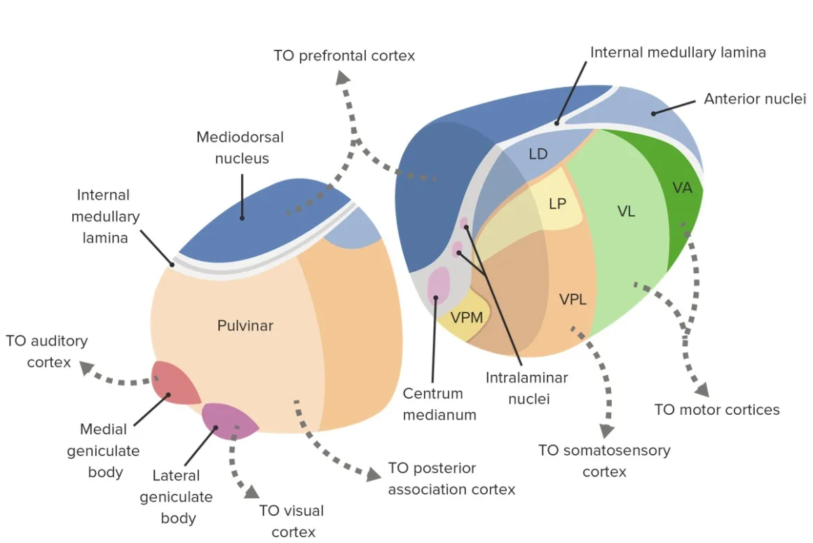
Anatomists typically group thalamic nuclei into five major functional categories. Sensory relay nuclei, including the ventral posterolateral and ventral posteromedial nuclei, handle touch, pain, and temperature from the body and face. The lateral and medial geniculate nuclei relay visual and auditory information respectively. Motor nuclei, such as the ventral anterior and ventral lateral nuclei, relay signals from the cerebellum and basal ganglia toward the motor cortex. Associative nuclei, including the pulvinar and the dorsomedial nucleus, support higher cognitive functions like attention and language. Finally, the intralaminar and reticular nuclei regulate arousal, alertness, and the overall flow of information through the thalamus itself.
- The ventral posterolateral nucleus relays touch, pain, and temperature signals from the body.
- The lateral geniculate nucleus relays visual information from the retina to the primary visual cortex.
- The medial geniculate nucleus relays auditory signals toward the primary auditory cortex.
- The reticular nucleus forms a thin shell around the thalamus and regulates arousal and thalamocortical rhythm.
Neuroscientist Rodolfo Llinás’s extensive research on thalamocortical circuitry demonstrated that these nuclei do not simply pass information along passively; they actively filter, synchronize, and rhythmically gate signals before the cortex ever receives them, a process central to his influential theory of thalamocortical dysrhythmia, which links disrupted thalamic rhythms to conditions ranging from chronic pain to certain psychiatric symptoms.
How Does the Thalamus Relay Sensory Information?
The thalamus acts as the mandatory checkpoint for nearly every sensory signal traveling toward conscious awareness. Vision, hearing, touch, taste, and balance all pass through specific thalamic nuclei before reaching the corresponding area of the cerebral cortex.
This relay function is far more active than the term “relay” might suggest. Rather than simply forwarding raw data, the thalamus performs real-time sensory gating, amplifying signals that seem important and dampening those judged irrelevant, which is part of how you can tune out a ticking clock but instantly notice your name spoken across a crowded room. Only olfactory information bypasses this thalamic checkpoint, traveling instead directly to the olfactory cortex, a notable exception that reflects smell’s ancient evolutionary pathway compared to the other senses.
Because each sensory modality is routed through its own dedicated nucleus, damage to a specific thalamic region tends to produce a correspondingly specific deficit. A lesion affecting the ventral posterolateral nucleus, for instance, can cause loss of touch and pain sensation confined to one side of the body, while damage to the lateral geniculate nucleus produces a distinctive visual field defect, even though the eyes and visual cortex themselves remain completely intact.
This precision makes the thalamus an important diagnostic landmark: when a patient reports one-sided numbness or vision loss that doesn’t match a typical cortical or peripheral nerve pattern, clinicians often consider a thalamic lesion as a strong possibility.
What Role Does the Thalamus Play in Motor Coordination?
Beyond sensory relay, the thalamus is a critical waypoint for motor signals moving between the cerebellum, basal ganglia, and cerebral cortex. Nearly every voluntary movement you make depends on thalamic circuits working correctly behind the scenes.
The ventral anterior and ventral lateral nuclei receive input from the cerebellum and basal ganglia, structures responsible for coordinating movement timing, smoothness, and initiation, and then relay this processed information to the primary motor cortex for final execution. This circuit explains why disorders affecting the basal ganglia, such as Parkinson’s disease, produce motor symptoms that trace back partly to disrupted thalamic relay function, even though the basal ganglia themselves are the primary site of pathology.
Neurosurgeon Wilder Penfield’s early work mapping brain function through direct stimulation helped establish just how tightly integrated the thalamus is with the cortical motor system, showing that thalamic circuits are not a passive stopover but an active participant in shaping how movement commands are refined before execution. This integration is part of why certain thalamic strokes produce a distinctive movement pattern sometimes called thalamic ataxia, marked by clumsy, poorly coordinated limb movements despite normal muscle strength.
For patients recovering from a thalamic stroke, this connection underscores why physical and occupational therapy targeting coordination and fine motor control, not just strength, forms a core part of effective rehabilitation.
How Does the Thalamus Regulate Sleep, Wakefulness, and Consciousness?
The thalamus plays an essential role in determining whether you are awake, drowsy, or deeply asleep, working in close partnership with the brainstem’s reticular activating system. Its rhythmic electrical activity essentially sets the tempo for different states of consciousness.
During deep, non-REM sleep, thalamic neurons shift into a distinctive oscillating firing pattern that effectively blocks most incoming sensory information from reaching the cortex, which is part of why you can sleep through moderate background noise but still wake instantly to a smoke alarm or your name being called. Sleep researcher Allan Hobson’s influential work on the neurobiology of dreaming highlighted how shifts in brainstem and thalamic activity during REM sleep allow vivid, often bizarre dream imagery to form, even while the thalamus continues gating which sensory signals from the outside world are allowed to intrude on the dreaming brain.
The thalamus’s role extends into baseline consciousness itself. Neuroscientist Giulio Tononi’s integrated information theory of consciousness identifies thalamocortical interactions as central to how the brain generates a unified, coherent conscious experience out of countless individual signals, suggesting that damage severe enough to disrupt this thalamocortical loop can produce profound alterations in awareness, ranging from mild confusion to coma, depending on the extent of injury.
A practical takeaway for anyone managing chronic sleep disruption: because thalamic rhythms are so central to the sleep-wake cycle, consistent sleep hygiene practices, like a stable bedtime and reduced late-night screen stimulation, work in part by supporting the thalamus’s natural gating rhythms rather than fighting against them.

How Is the Thalamus Involved in Emotion and Memory?
The thalamus is far more than a sensory switchboard; it also participates directly in emotional processing and memory formation through its dense connections with the limbic system. This involvement explains why thalamic damage can sometimes produce emotional changes that seem disconnected from any obvious sensory or motor symptom.
Certain thalamic nuclei, particularly the anterior and dorsomedial nuclei, connect closely with the hippocampus, mammillary bodies, and fornix, structures central to forming new episodic memories. Damage to these specific thalamic regions, sometimes from stroke or chronic alcohol-related thiamine deficiency, can produce a form of amnesia strikingly similar to that seen with hippocampal damage, underscoring that memory formation depends on an entire interconnected circuit rather than any single structure working in isolation.
The thalamus also plays a role in fear processing through a rapid, subcortical pathway to the amygdala. Neuroscientist Joseph LeDoux’s research on fear conditioning demonstrated that sensory information can travel a fast “low road” directly from the thalamus to the amygdala, triggering an instant threat response before the slower, more detailed “high road” through the cortex has finished analyzing the situation. This dual-pathway system is why you might flinch at a shadow before consciously recognizing it as harmless, a survival-oriented shortcut that prioritizes speed over accuracy in the earliest milliseconds of threat detection.
Physician and neuroscientist Antonio Damasio’s broader work on the neural basis of consciousness and emotion has emphasized how deeply intertwined bodily feeling states, thalamic relay, and cortical awareness are in constructing a coherent sense of self, reinforcing that the thalamus is woven into far more than pure sensation.
What Happens When the Thalamus Is Damaged?
Thalamic damage produces an unusually wide and sometimes puzzling range of symptoms, precisely because so many distinct functions are packed into such a small structure. The exact presentation depends heavily on which specific nucleus or nuclei are affected.
Stroke is the most common cause of thalamic injury, often producing a striking and distinctive pattern called thalamic pain syndrome (also known as Dejerine-Roussy syndrome), in which a person develops persistent, often severe burning or aching pain on one side of the body, sometimes appearing weeks after the initial stroke and after sensation has partially recovered. Other thalamic lesions can cause profound sensory loss, movement incoordination, disturbed consciousness ranging from drowsiness to coma, or, when the anterior and dorsomedial nuclei are involved, significant memory impairment resembling other forms of amnesia.
- Thalamic pain syndrome produces persistent burning or aching pain following thalamic stroke, sometimes with a delayed onset.
- Sensory loss confined to one side of the body can occur with damage to specific relay nuclei.
- Thalamic amnesia resembles hippocampal memory loss when the anterior or dorsomedial nuclei are affected.
- Altered consciousness, from mild confusion to coma, can result from more extensive bilateral thalamic injury.
Because these symptoms can appear so varied and sometimes contradictory, from numbness to pain to memory loss, thalamic strokes are occasionally misattributed to psychiatric causes before imaging reveals the true underlying neurological explanation, which is an important reason to pursue thorough medical evaluation for any sudden, unexplained combination of sensory and cognitive changes.
How Are Thalamic Conditions Diagnosed and Treated?
Diagnosis of thalamic dysfunction typically begins with a detailed neurological examination assessing sensation, movement, alertness, and memory, since the specific combination of deficits can help localize the problem before any scan is performed. From there, targeted imaging and, when appropriate, electrophysiological testing confirm the diagnosis.
MRI is the preferred imaging modality for visualizing the thalamus and identifying strokes, tumors, or other structural abnormalities, since its deep, central location can make certain lesions harder to detect on standard CT imaging. Electroencephalography can help assess disruptions in thalamocortical rhythm relevant to consciousness or sleep disorders, while formal neuropsychological testing helps characterize the specific pattern of memory or cognitive impairment when thalamic amnesia is suspected.
Treatment depends entirely on the underlying cause. Thalamic pain syndrome is notoriously difficult to treat and often requires a combination of specific neuropathic pain medications rather than standard painkillers, since typical analgesics frequently provide limited relief for this particular type of nerve-related pain. Rehabilitation for thalamic stroke commonly combines physical therapy for coordination, occupational therapy for daily functioning, and, when memory is affected, structured cognitive rehabilitation strategies.
- Seek prompt evaluation for any sudden combination of numbness, weakness, or confusion, since thalamic stroke requires urgent treatment.
- Report delayed pain that develops weeks after a stroke, since thalamic pain syndrome can emerge well after the initial event.
- Request specialized neuropathic pain treatment rather than standard painkillers if thalamic pain is suspected.
- Pursue structured cognitive rehabilitation promptly when memory changes accompany thalamic injury, since early intervention supports better functional outcomes.
How Does Thalamic Function Change Across the Lifespan?
The thalamus develops early and continues refining its connections throughout childhood, while also showing measurable vulnerability to specific conditions in older adulthood. Recognizing this trajectory helps clarify what counts as normal development versus a pattern that warrants medical attention.
In fetal and early childhood development, the thalamus forms one of the earliest functioning relay systems in the brain, with thalamocortical connections beginning to establish themselves well before birth and continuing to refine into adolescence as sensory processing becomes increasingly precise. Disruptions to this early thalamocortical development have been studied in relation to certain neurodevelopmental conditions, since accurate sensory gating and filtering depend on these circuits maturing correctly.
In older adults, cardiovascular risk factors such as hypertension and diabetes increase vulnerability to small perforating-artery strokes affecting the thalamus, given how many of its blood vessels are narrow and prone to blockage. Certain neurodegenerative conditions, including some forms of dementia, also involve measurable thalamic volume loss, contributing to the sleep disturbances and attentional difficulties commonly seen in these conditions. Fatal familial insomnia, a rare genetic prion disease, offers a striking, if severe, illustration of just how central the thalamus is to sleep regulation, since progressive damage to specific thalamic nuclei in this condition produces relentless, ultimately fatal insomnia.
Supporting healthy thalamic function across the lifespan includes managing cardiovascular risk factors, prioritizing consistent sleep patterns, and seeking prompt medical attention for any sudden, unexplained combination of sensory, motor, or cognitive changes rather than assuming they will resolve on their own.
FAQs about the Thalamus
What is the main function of the thalamus?
The thalamus’s primary role is acting as the brain’s central relay station, processing and forwarding nearly all sensory and motor information between the body, brainstem, and cerebral cortex. Beyond simple relay, it actively filters and prioritizes signals through a process called sensory gating, deciding what reaches conscious awareness and what gets suppressed. It also contributes to sleep-wake regulation, emotional processing, and memory formation through its dense connections with the limbic system. This combination of functions is why thalamic damage can produce such a wide and sometimes puzzling variety of symptoms.
Where exactly is the thalamus located in the brain?
The thalamus sits at the very center of the brain, within a region called the diencephalon, positioned just above the brainstem and lateral to the third ventricle. It consists of two egg-shaped masses, one in each hemisphere, connected across the midline by a bridge of tissue called the interthalamic adhesion. This central location allows it to send nerve fiber connections outward in all directions toward the cerebral cortex, forming the thalamocortical radiations essential to its relay function. Its deep, centrally embedded position also means damage here tends to affect multiple functions simultaneously rather than a single isolated ability.
What happens if the thalamus is damaged?
Thalamic damage can cause a wide range of symptoms depending on which specific nuclei are affected, including one-sided sensory loss, movement incoordination, disrupted consciousness, and memory impairment. A particularly distinctive outcome is thalamic pain syndrome, where persistent burning or aching pain develops on one side of the body, sometimes weeks after a stroke. Because so many distinct functions are packed into this small structure, symptoms can appear unusually varied or even contradictory, occasionally leading to initial misdiagnosis before imaging clarifies the underlying thalamic cause. Prompt neurological evaluation is important for any sudden combination of these symptoms.
How does the thalamus affect sleep?
The thalamus plays a central role in regulating the sleep-wake cycle by shifting its electrical activity between distinct patterns for wakefulness, deep sleep, and REM sleep. During deep sleep, thalamic neurons largely block incoming sensory information from reaching the cortex, which is part of why moderate background noise doesn’t wake most sleepers while more significant or personally relevant sounds still do. Disruptions to thalamic rhythms have been linked to various sleep disturbances, and in rare genetic conditions like fatal familial insomnia, progressive damage to specific thalamic nuclei severely disrupts the ability to sleep altogether. Supporting healthy sleep patterns through consistent routines helps reinforce these natural thalamic rhythms.
Can thalamus damage cause memory loss?
Yes, particularly when damage affects the anterior or dorsomedial thalamic nuclei, which connect closely with the hippocampus and other memory-related limbic structures. This type of injury can produce a form of amnesia that closely resembles memory loss caused by direct hippocampal damage, since memory formation relies on an interconnected circuit rather than any single brain region working alone. Common causes include stroke and chronic thiamine deficiency related to alcohol use, both of which can selectively affect these vulnerable thalamic regions. Structured cognitive rehabilitation can help some patients develop compensatory memory strategies, even when the underlying impairment does not fully resolve.
Is thalamic pain syndrome treatable?
Thalamic pain syndrome, also called Dejerine-Roussy syndrome, is notoriously challenging to treat, but meaningful symptom relief is achievable for many patients with the right approach. Standard over-the-counter pain relievers typically provide limited benefit, since this pain arises from disrupted nerve signaling rather than tissue injury, so treatment usually relies on specific neuropathic pain medications, and sometimes additional interventions like nerve stimulation therapies for cases resistant to medication. Because the pain can emerge weeks after the initial stroke and may worsen over time, ongoing communication with a neurologist or pain specialist is important for adjusting treatment as symptoms evolve. Multidisciplinary pain management, including psychological support, often improves quality of life alongside medical treatment.
Bibliography
- Torrico, T. J., & Munakomi, S. (2023). Neuroanatomy, Thalamus. StatPearls Publishing, National Center for Biotechnology Information Bookshelf.
- Cleveland Clinic. (2022). Thalamus: What It Is, Function, Location & Disorders.
- Kenhub. (2015). Thalamus: Anatomy, Nuclei, Function.
- Physio-pedia. (2025). Thalamus.
- Llinás, R. R., & Ribary, U. (2001). Consciousness and the brain: the thalamocortical dialogue in health and disease. Annals of the New York Academy of Sciences.
- Hobson, J. A. (1988). The Dreaming Brain. Basic Books.
- Tononi, G., & Koch, C. (2015). Consciousness: Here, there and everywhere? Philosophical Transactions of the Royal Society B.
- LeDoux, J. E. (1996). The Emotional Brain: The Mysterious Underpinnings of Emotional Life. Simon & Schuster.
- Damasio, A. R. (1999). The Feeling of What Happens: Body and Emotion in the Making of Consciousness. Harcourt Brace.
- Penfield, W., & Jasper, H. (1954). Epilepsy and the Functional Anatomy of the Human Brain. Little, Brown and Company.
Use this citation format to reference the article clearly and help readers find the original source.
PsychologyFor. (2026). Thalamus: What it Is, Functions and Location. PsychologyFor. https://psychologyfor.com/thalamus-what-it-is-functions-and-location/