


See more of our articles in your search results.
Picture the human body as a house you’ve lived in your whole life, yet you’ve never actually seen inside the walls, never traced where the pipes run or how the wiring connects the switch by the door to the light three rooms away. That’s roughly how most people relate to their own anatomy: aware that something intricate is happening beneath the skin, but with no real map of how it all fits together. If you’ve ever wondered why a cardiologist and an embryologist study “the same” heart in completely different ways, or why a textbook diagram of a kidney looks nothing like what a surgeon actually sees during an operation, you’re bumping into the fact that anatomy is not one single discipline but a family of related fields, each with its own tools, scale, and purpose.
This distinction matters more than it might first appear, especially if you’re a student trying to make sense of a curriculum that suddenly splits “anatomy” into gross anatomy, histology, embryology, and other seemingly separate subjects. Without understanding why these branches exist, it’s easy to feel like you’re drowning in disconnected facts: bone names in one class, cell structures in another, embryonic folding in a third, with no clear thread tying them together. In reality, each branch simply looks at the same body through a different lens, whether that’s the naked eye, a microscope, a developmental timeline, or a cross-species comparison, and knowing which lens you’re using at any given moment makes the material dramatically easier to organize and retain. This is especially true for students in medicine, nursing, physical therapy, and biology, where confusing one branch’s methods with another’s can lead to real gaps in clinical reasoning later on.
So how many branches are there, and what exactly separates one from the next?
This article walks through the 5 branches of anatomy, explaining what each one studies, the tools and techniques associated with it, and how they all connect to form a complete picture of the human body.
What Is Anatomy and Why Is It Divided Into Branches?
Anatomy is the branch of biological science dedicated to studying the structure of organisms and the relationships between their parts. Because the human body can be examined at wildly different scales, from an entire organ system down to a single cell, and from a fully formed adult back to a two-week-old embryo, anatomists have organized their field into distinct branches, each with a specific focus, methodology, and set of tools.
How many of these have you been "meaning to read" for months?
- 1
 TarcherThe Artist's Way30th Anniversary EditionListen Free On Audible
 - 2
 New Harbinger PublicationsThe Dialectical Behavior Therapy Skills WorkbookPractical DBT Exercises for Learning Mindfulness, Interpersonal Effectiveness, Emotion Regulation...Listen Free On Audible
 - 3
 New Harbinger PublicationsAdult Children of Emotionally Immature ParentsHow to Heal from Distant, Rejecting, or Self-Involved ParentsListen Free On Audible
This division isn’t arbitrary. The Renaissance anatomist Andreas Vesalius, often called the father of modern human anatomy, transformed the field in the sixteenth century by insisting on direct human dissection rather than relying solely on ancient texts, and his meticulous, hands-on approach in works like De Humani Corporis Fabrica essentially established gross anatomy as a rigorous discipline separate from philosophical speculation about the body. As microscopes, embryological staining techniques, and comparative zoological studies developed over the following centuries, new branches naturally split off to handle what gross dissection alone couldn’t reveal.
Today, most anatomy curricula recognize five major branches:
- Gross anatomy, the study of structures visible to the naked eye.
- Microscopic anatomy, the study of tissues and cells under magnification.
- Developmental anatomy, the study of how the body forms and changes from conception through maturity.
- Surface anatomy, the study of external body features and their relationship to internal structures.
- Comparative anatomy, the study of structural similarities and differences across species.
Each of these branches uses different tools and reference points, yet all of them ultimately serve the same goal: producing an accurate, functional understanding of how living structures are organized.
What Is Gross Anatomy (Macroscopic Anatomy)?
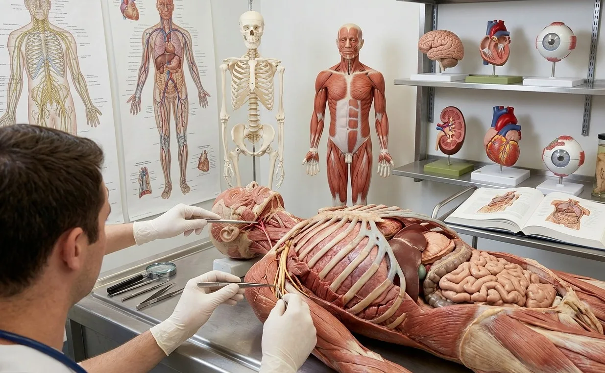
Gross anatomy, also called macroscopic anatomy, is the study of body structures large enough to be seen without magnification, typically through dissection, palpation, and imaging. It forms the backbone of most medical and biological education because it maps directly onto what clinicians encounter during physical exams and surgery.
Gross anatomy itself is commonly studied through two complementary approaches. Regional anatomy examines all the structures within a specific area of the body at once, such as everything located in the thorax or the upper limb, which mirrors how surgeons think when operating on a particular region. Systemic anatomy, by contrast, studies one organ system at a time across the entire body, such as tracing the cardiovascular system from the heart through every vessel, which is often how anatomy is first taught to students because it builds a functional understanding of each system before layering in regional complexity.
Gross anatomy relies heavily on cadaveric dissection, a tradition with roots stretching back to Vesalius’s public dissections in Padua, as well as modern medical imaging techniques like X-ray, CT, and MRI, which allow clinicians to visualize gross structures in living patients without invasive procedures. The nineteenth-century anatomist Henry Gray, whose name lives on through the enduring reference text Gray’s Anatomy, helped standardize how gross anatomical knowledge was documented and taught, and his detailed illustrations remain foundational to how the subject is presented even in contemporary editions.
Because gross anatomy deals with structures visible to the eye, it is the branch most directly applied in fields like surgery, physical therapy, radiology, and emergency medicine, where practitioners need to quickly locate and assess organs, muscles, nerves, and vessels in real time.
What Is Microscopic Anatomy (Histology and Cytology)?
Microscopic anatomy studies structures too small to be seen with the naked eye, primarily tissues and cells, using microscopes and specialized staining techniques. It splits into two closely related subfields: histology, which examines tissue organization, and cytology, which focuses specifically on the internal structure and function of individual cells.
The foundations of this branch trace back to the seventeenth-century Italian physician Marcello Malpighi, frequently regarded as the founder of microscopic anatomy, whose pioneering use of early microscopes revealed structures like capillaries and the layered organization of skin that were completely invisible to earlier anatomists working only with dissection. His discoveries proved that the body’s macroscopic organs were themselves built from smaller, highly organized structural units, a realization that opened an entirely new dimension of anatomical inquiry.
Modern microscopic anatomy depends on techniques largely unavailable in Malpighi’s era, including thin tissue sectioning, chemical staining that highlights specific cellular components, and increasingly sophisticated tools like electron microscopy, which can resolve structures at the nanometer scale. This branch is essential for understanding:
- How the four basic tissue types (epithelial, connective, muscle, and nervous) are organized and function.
- How disease processes alter normal cellular architecture, which underlies most diagnostic pathology.
- How organs are constructed from specialized tissue layers working together.
Pathologists rely heavily on microscopic anatomy to diagnose disease from tissue biopsies, since many conditions, including most cancers, are identified by characteristic changes at the cellular level long before they produce visible symptoms or gross structural changes.
What Is Developmental Anatomy (Embryology)?
Developmental anatomy, most commonly known as embryology, studies how the body’s structures form, differentiate, and change from fertilization through birth and, in a broader sense, throughout the entire human lifespan. This branch explains not just what a structure looks like in its final adult form, but how it came to exist in the first place.
The nineteenth-century German anatomist Wilhelm His made significant early contributions to this field by carefully documenting the physical folding and layering processes that transform a simple embryonic disc into the complex, three-dimensional structures of a developing fetus, work that helped establish embryology as a rigorous descriptive science rather than speculative theory. Developmental anatomy typically divides into prenatal development, covering the dramatic structural changes from fertilization through the embryonic and fetal periods, and postnatal development, which tracks growth and structural maturation from birth through adulthood and aging.
Understanding developmental anatomy has significant clinical relevance, because many congenital conditions, such as heart defects or neural tube abnormalities, arise from specific disruptions during defined windows of embryonic development. Knowing exactly when and how a structure normally forms allows clinicians to understand why certain developmental errors produce particular, predictable patterns of malformation rather than random ones.
This branch also has significant educational value beyond medicine, since tracing how a structure develops often makes its adult anatomy far more intuitive. For example, understanding how the heart forms from a simple folded tube helps explain why its adult chambers and major vessels are arranged the way they are, rather than requiring rote memorization of a seemingly arbitrary final layout.

What Is Surface Anatomy?
Surface anatomy is the study of external body features and how they correspond to the location of internal structures lying beneath the skin. It essentially functions as a practical bridge between what a clinician can see and feel from the outside and what is actually happening inside the body.
This branch relies on visible or palpable landmarks, such as bony prominences, skin creases, and muscle contours, to estimate the position of deeper structures without needing imaging or dissection. A physician locating the apex of the heart by counting rib spaces, or a nurse identifying a vein for blood draw based on visible surface contours, is applying surface anatomy directly in a clinical setting.
Surface anatomy is particularly valuable in emergency and outpatient settings, where quick, accurate physical assessment matters more than detailed imaging. Common applications include:
- Identifying pulse points for cardiovascular assessment.
- Locating injection or blood draw sites using visible venous landmarks.
- Estimating organ borders for physical examination, such as percussing liver or lung boundaries.
- Guiding safe placement of needles or catheters relative to nearby nerves and vessels.
Because surface anatomy depends on visible and palpable cues, it requires strong integration with gross anatomical knowledge; clinicians must know not just what a landmark looks like, but what structure it corresponds to and how variable that relationship can be between individuals.
What Is Comparative Anatomy?
Comparative anatomy studies the similarities and differences in body structures across different species, using those comparisons to understand evolutionary relationships, functional adaptations, and the underlying principles of biological design. Rather than focusing on the human body alone, this branch places human anatomy within the broader context of the animal kingdom.
The French naturalist Georges Cuvier, working in the late eighteenth and early nineteenth centuries, is widely credited with establishing comparative anatomy as a systematic scientific discipline. Cuvier famously demonstrated that an animal’s anatomical structures were so functionally interconnected that a skilled comparative anatomist could infer an entire animal’s likely appearance and habits from a single bone, a principle he called the “correlation of parts.” His work laid essential groundwork for later evolutionary biology, since consistent structural patterns across species provided some of the earliest evidence for common ancestry.
Comparative anatomy remains highly relevant today, particularly in these areas:
- Evolutionary biology, where shared structures across species (homologous structures) reveal common ancestry.
- Biomedical research, where animal models are chosen based on anatomical and physiological similarity to humans.
- Veterinary medicine, which depends entirely on understanding structural variation across different animal species.
- Paleontology, where comparative anatomical principles help reconstruct extinct species from fossilized remains.
This branch also deepens understanding of human anatomy itself, since recognizing which human structures are shared broadly across vertebrates, and which are uniquely specialized, clarifies which features are evolutionarily ancient versus recently adapted.

How Do the 5 Branches of Anatomy Work Together?
The five branches of anatomy are not isolated silos; they function as complementary lenses that, combined, produce a far richer understanding than any single branch could offer alone. A single organ, such as the heart, can be fully appreciated only by integrating knowledge from all five perspectives at once.
Consider how this plays out in practice: gross anatomy describes the heart’s four chambers and major vessels as seen during dissection or surgery; microscopic anatomy reveals the specialized cardiac muscle tissue and conduction fibers that allow coordinated contraction; developmental anatomy explains how that four-chambered structure formed from a simple embryonic tube through a precise folding and septation process; surface anatomy guides clinicians in locating the heart’s borders and auscultation points on a living patient’s chest; and comparative anatomy shows how the four-chambered mammalian heart differs from the three-chambered hearts found in most reptiles, illuminating functional trade-offs in circulatory efficiency.
This integrated approach explains why anatomy curricula, particularly in health-related professions, deliberately weave multiple branches together rather than teaching them in complete isolation. A clinician who only understands gross anatomy might correctly identify an organ’s location but miss why a particular developmental anomaly produces a specific structural defect, while a researcher who only understands microscopic anatomy might miss how cellular-level findings translate into whole-body clinical symptoms.
| Branch | Primary Focus |
|---|---|
| Gross anatomy | Structures visible without magnification, studied by region or system |
| Microscopic anatomy | Tissues (histology) and cells (cytology) studied under magnification |
| Developmental anatomy | Formation and structural change from embryo through adulthood |
| Surface anatomy | External landmarks correlated with underlying internal structures |
| Comparative anatomy | Structural similarities and differences across species |
Why Does Understanding These Branches Matter for Students and Professionals?
Recognizing which branch of anatomy a given topic belongs to helps students organize study material far more effectively than treating “anatomy” as one undifferentiated subject. When a lecture shifts from discussing bone names to discussing bone cell types, that’s a shift from gross to microscopic anatomy, and recognizing that shift helps a student anticipate what kind of information, structural naming versus cellular function, they should expect to retain and apply.
For professionals, this framework directly shapes daily practice. A surgeon depends primarily on gross and surface anatomy to navigate safely during an operation, while a pathologist depends primarily on microscopic anatomy to diagnose disease from tissue samples. A pediatric specialist treating a congenital anomaly draws heavily on developmental anatomy to understand why a particular malformation occurred, while a researcher developing a new animal model for drug testing relies on comparative anatomy to select a species with sufficiently similar physiology to humans. Recognizing which branch applies to a given clinical or research question ensures that the right tools, terminology, and reference frameworks are used from the outset, reducing the risk of miscommunication or diagnostic error.
Beyond formal professional applications, understanding these branches also helps anyone reading general health information make sense of why certain claims come from certain types of evidence. A claim about “what a healthy artery looks like on imaging” draws on gross anatomy, while a claim about “what happens to artery walls at the cellular level in atherosclerosis” draws on microscopic anatomy, and recognizing that distinction helps readers evaluate health information with more critical clarity.
FAQs About the Branches of Anatomy
What are the 5 branches of anatomy?
The five widely recognized branches of anatomy are gross anatomy, microscopic anatomy, developmental anatomy, surface anatomy, and comparative anatomy. Gross anatomy studies structures visible without magnification, microscopic anatomy examines tissues and cells under magnification, and developmental anatomy traces how body structures form and change over time, particularly during embryonic development. Surface anatomy connects external, visible body landmarks to underlying internal structures, which is especially useful in physical examinations, while comparative anatomy studies structural similarities and differences across different animal species. Together, these branches provide a complete anatomical framework, covering everything from whole organs down to individual cells, and from a single human body to patterns across the entire animal kingdom.
What is the difference between gross anatomy and microscopic anatomy?
Gross anatomy studies structures large enough to see without any magnification, such as organs, bones, and muscles, typically through dissection, palpation, or medical imaging. Microscopic anatomy, by contrast, studies structures that require magnification to observe, primarily tissues (the focus of histology) and individual cells (the focus of cytology). The two branches are complementary rather than competing: gross anatomy tells you what an organ looks like and where it sits in the body, while microscopic anatomy explains how that organ is actually built and functions at a cellular level. Medical and biology curricula typically teach both together, since a full understanding of any organ requires both scales of observation working in tandem.
Why is embryology considered a branch of anatomy?
Embryology, more formally called developmental anatomy, is considered a branch of anatomy because it directly explains the structural origins of the body parts studied in other anatomical branches. Rather than describing only the finished, adult form of a structure, developmental anatomy traces how that structure came into existence, which folding, differentiation, and growth processes produced its final shape and position. This is particularly important in clinical contexts, since understanding normal development helps explain why certain congenital conditions produce specific, predictable structural abnormalities rather than random ones. Many anatomy programs integrate embryology directly alongside gross anatomy instruction, since knowing how a structure formed often makes its adult anatomy far easier to understand and remember.
How is surface anatomy used in clinical practice?
Surface anatomy is used constantly in clinical practice to locate internal structures without needing imaging or invasive procedures, relying instead on visible or palpable external landmarks. Physicians use surface landmarks to find pulse points, estimate organ boundaries during physical examination, identify safe sites for injections or blood draws, and guide procedures like inserting central lines near major blood vessels. This branch is especially valuable in emergency situations, where quick, accurate physical assessment can be lifesaving before diagnostic imaging is available. Strong surface anatomy knowledge essentially allows clinicians to build a reliable internal map of the body using only what they can see and feel from the outside.
What is the purpose of comparative anatomy?
Comparative anatomy exists to study structural similarities and differences across different animal species, using those patterns to understand evolutionary relationships, functional adaptations, and broader biological principles. This branch has practical applications well beyond pure biology, including guiding the selection of appropriate animal models for biomedical research, informing veterinary medicine across diverse species, and helping paleontologists reconstruct extinct organisms from incomplete fossil remains. Comparative anatomy also enriches human anatomical understanding, since recognizing which structures are shared broadly across the animal kingdom, known as homologous structures, versus which are uniquely human helps clarify which anatomical features are evolutionarily ancient and which are more recently specialized. This branch essentially places human anatomy in context rather than treating it as an isolated case.
Do all anatomy courses cover all 5 branches equally?
No, the emphasis given to each branch varies significantly depending on the academic program and professional goal. Medical, nursing, and physical therapy programs typically emphasize gross anatomy and surface anatomy heavily, since these directly support physical examination, diagnostic reasoning, and procedural skills, while dedicating comparatively less time to comparative anatomy. Biology and evolutionary science programs, on the other hand, often place much greater emphasis on comparative anatomy, since understanding structural variation across species is central to that field. Pathology and cell biology tracks understandably prioritize microscopic anatomy, while pediatric and genetics-focused tracks tend to give developmental anatomy more weight. This variation reflects the fact that each branch serves different practical and academic priorities depending on a student’s eventual specialization.
Bibliography
- Vesalius, A. (1543). De Humani Corporis Fabrica Libri Septem.
- Gray, H. (1858). Gray’s Anatomy: Descriptive and Surgical.
- Standring, S. (Ed.). (2020). Gray’s Anatomy: The Anatomical Basis of Clinical Practice. Elsevier.
- Moore, K. L., Dalley, A. F., & Agur, A. M. R. (2018). Clinically Oriented Anatomy. Wolters Kluwer.
- Sadler, T. W. (2019). Langman’s Medical Embryology. Wolters Kluwer.
- Cuvier, G. (1800). Leçons d’anatomie comparée.
- Mescher, A. L. (2021). Junqueira’s Basic Histology: Text and Atlas. McGraw Hill.
- His, W. (1885). Anatomie menschlicher Embryonen.
Use this citation format to reference the article clearly and help readers find the original source.
PsychologyFor. (2026). The 5 Branches of Anatomy (And Their Characteristics). PsychologyFor. https://psychologyfor.com/the-5-branches-of-anatomy-and-their-characteristics/