

See more of our articles in your search results.
You sit down to make a decision that should be simple — what to eat, whether to speak up in a meeting, how to respond to a difficult text message — and your mind suddenly feels like it’s pulling in five directions at once. Or you watch someone you love recover from a brain injury and struggle with something as basic as finding the right word, planning their day, or reading someone’s facial expression, and you wonder how one injury could touch so many different parts of who they are. These moments, confusing as they feel, all trace back to the most complex, densely packed structure in the human body: the cerebral cortex.
It can feel overwhelming to realize how much of your identity — your personality, your judgment, your ability to plan, speak, and connect with others — depends on a thin sheet of tissue barely a few millimeters thick. This becomes especially disorienting for people navigating a new diagnosis, a brain injury, or simply the ordinary experience of forgetfulness and mental fatigue, since it’s hard to locate exactly where these experiences “live” in the body the way you might locate a sore muscle. Understanding the structure and function of this remarkable outer layer of the brain can replace confusion with a clearer, more compassionate sense of how thought, emotion, and behavior actually work. It also helps explain why certain symptoms cluster together in very specific, predictable patterns.
So what exactly is happening in this thin, folded layer that makes it responsible for nearly everything we consider distinctly human?
This guide breaks down the anatomy, layers, lobes, and everyday functions of the cerebral cortex, along with what happens when different parts of it are injured or impaired.
What Is the Cerebral Cortex and Where Is It Located?
The cerebral cortex is the outermost layer of the brain, a thin, deeply folded sheet of gray matter that covers the two cerebral hemispheres. It is the seat of most higher-order mental activity, including conscious thought, perception, language, and voluntary movement, making it arguably the most important structure for what we experience as our sense of self.
How many of these have you been "meaning to read" for months?
- 1
 Sounds TrueHow to Carry What Can't Be FixedA Journal for GriefListen Free On Audible
 - 2
 AzujosThe Zen Monkey and the Lotus Flower52 Stories to Relieve Stress, Stop Negative Thoughts, Find Happiness, and Live Your Best LifeListen Free On Audible
 - 3
 MajostaDon't Believe Everything You ThinkWhy Your Thinking Is The Beginning & End Of Suffering (Beyond Suffering)Listen Free On Audible
Physically, the cerebral cortex ranges from about one and a half to four and a half millimeters thick, yet if it were unfolded and flattened out, it would cover an area roughly the size of a large dinner napkin. This is only possible because of its distinctive folding pattern, made up of raised ridges called gyri and deep grooves called sulci, an evolutionary adaptation that allows an enormous surface area of neural tissue to fit within the limited space of the skull.
Beneath this thin gray matter layer lies the brain’s white matter, made up of myelinated nerve fibers that connect different cortical regions to each other and to deeper brain structures. The cerebral cortex itself contains an estimated fourteen to sixteen billion neurons, organized into an intricate, six-layered structure that varies subtly in thickness and cell composition depending on the specific function of that region.
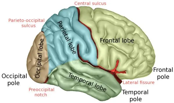
Major landmarks help divide the cortex into recognizable regions: the central sulcus separates the frontal and parietal lobes, the lateral or Sylvian fissure separates the temporal lobe from the frontal and parietal lobes above it, and the parieto-occipital sulcus marks the boundary between the parietal and occipital lobes. These divisions form the basis for understanding the cortex’s four major cerebral lobes, each specialized for a distinct set of functions.
What Are the Layers of the Cerebral Cortex?
Most of the cerebral cortex is organized into six distinct horizontal layers, each with a characteristic mix of neuron types and connection patterns. This layered architecture, known as the neocortex, is one of the defining features that distinguishes the human brain’s information-processing capacity.
Layer one, closest to the brain’s surface, contains relatively few neuron cell bodies and instead consists mostly of connecting fibers. Layers two and three contain neurons that primarily connect to other cortical areas, supporting communication across different regions of the brain. Layer four is the primary destination for incoming sensory information arriving from the thalamus, making it especially prominent in sensory processing regions like the visual and auditory cortex. Layers five and six send output signals downward to subcortical structures, the brainstem, and the spinal cord, making them especially thick and prominent in motor regions like the primary motor cortex.
This layered organization is not uniform across the entire cortex; regions dedicated to different tasks show variations in the relative thickness of each layer, a pattern of variation historian and neuroanatomist Korbinian Brodmann famously mapped in the early twentieth century, dividing the cortex into numbered regions still referenced today, such as Brodmann area 4 for the primary motor cortex.
- The outer cortical layers primarily support communication between different cortical regions.
- The middle layer serves as the main entry point for sensory information from the thalamus.
- The deep cortical layers send output signals to subcortical structures and the spinal cord.
- Regional variation in this layered architecture corresponds closely to functional specialization across the cortex.

What Are the Four Lobes of the Cerebral Cortex and Their Functions?
The cerebral cortex is divided into four paired lobes — frontal, parietal, temporal, and occipital — each specializing in a different category of mental and physical function. Understanding this division offers a practical map for connecting specific symptoms to specific brain regions.
The frontal lobe, the largest of the four, sits at the front of the brain and governs voluntary movement, speech production, and much of what we consider executive function, including planning, judgment, and impulse control. The parietal lobe, positioned just behind it, processes touch, pressure, temperature, and pain, while also integrating this sensory information with vision to support spatial awareness and navigation. The temporal lobe, located along the sides of the brain near the ears, handles auditory processing, language comprehension, and aspects of memory formation. The occipital lobe, at the very back of the brain, is dedicated almost entirely to visual processing and interpretation.
| Lobe | Primary Functions |
| Frontal lobe | Voluntary movement, speech production, executive function, personality |
| Parietal lobe | Touch, spatial awareness, sensory integration |
| Temporal lobe | Hearing, language comprehension, memory |
| Occipital lobe | Visual processing and interpretation |
Some anatomists also recognize a fifth region, the limbic lobe, tucked along the inner surface of the hemispheres and involved in emotion, motivation, and memory, though it is often discussed separately from the four primary lobes due to its distinct evolutionary history and closer ties to subcortical structures like the amygdala and hippocampus.
How Do Sensory, Motor, and Association Areas Differ?
Beyond the four lobes, neuroscientists also classify the cerebral cortex functionally into three broad categories: sensory areas, motor areas, and association areas. This functional framework helps explain how raw information becomes meaningful experience and purposeful action.
Primary sensory areas receive and process incoming information from the senses, including the primary visual cortex in the occipital lobe, the primary auditory cortex in the temporal lobe, and the primary somatosensory cortex in the parietal lobe. Primary motor areas, concentrated in the frontal lobe’s precentral gyrus, generate the signals that produce voluntary movement. Neurosurgeon Wilder Penfield’s landmark research using direct cortical stimulation during awake brain surgery produced the famous cortical homunculus, a distorted map showing how different body parts are represented across the motor and sensory strips in proportion to how finely controlled or sensitive they need to be, rather than their actual size.
Association areas make up the majority of the cortex’s remaining surface and are responsible for interpreting, integrating, and giving meaning to raw sensory and motor information. These regions allow you to recognize a face as familiar, understand the emotional tone of someone’s voice, or plan a multi-step task, functions that go well beyond simple perception or movement. Association areas develop and mature more slowly than primary sensory and motor regions, which partly explains why complex reasoning and judgment continue developing well into early adulthood.
A practical takeaway: because association areas integrate information across multiple senses and brain regions, activities that combine sensory, motor, and cognitive engagement, like learning a new skill or hobby, tend to support broader cortical health more effectively than passive, single-sense activities alone.

How Do the Two Hemispheres of the Cerebral Cortex Differ?
The cerebral cortex is split into two hemispheres connected by a thick band of fibers called the corpus callosum, and while both sides handle similar categories of information, each shows distinct specializations. This division of labor is often referred to as hemispheric lateralization.
In most people, the left hemisphere shows greater specialization for sequential, analytical, and verbal processing, including most language production and comprehension functions. The right hemisphere tends toward more holistic, spatial, and simultaneous processing, contributing more heavily to recognizing faces, interpreting emotional tone, and understanding spatial relationships. Neuroscientist Michael Gazzaniga’s extensive research with split-brain patients, whose corpus callosum had been surgically severed to control severe epilepsy, provided some of the clearest evidence for this lateralization, revealing that each hemisphere can process information and even generate distinct responses somewhat independently when their communication is disrupted.
It’s worth noting that this popular idea of being purely “left-brained” or “right-brained” oversimplifies the reality; nearly every complex task recruits both hemispheres working together through the corpus callosum, and lateralization reflects a matter of relative specialization rather than strict, exclusive division. Damage confined to one hemisphere, however, can still produce a recognizable pattern of deficits consistent with that side’s typical strengths, which is clinically useful for localizing injury.
Understanding this lateralized specialization can help families make sense of stroke recovery patterns, since left-hemisphere damage more often affects language while right-hemisphere damage more often affects spatial awareness and emotional interpretation of speech.
How Does the Cerebral Cortex Support Language and Communication?
Language depends on a network of specialized cortical regions working in close coordination, primarily located in the dominant hemisphere. Damage to specific areas within this network produces strikingly different, highly specific language impairments.
Nineteenth-century physician Paul Broca identified a region in the frontal lobe, now called Broca’s area, essential for language production; damage here produces halting, effortful speech with relatively preserved comprehension, a pattern known as Broca’s aphasia. Around the same period, neurologist Carl Wernicke identified a separate region in the temporal lobe, now called Wernicke’s area, essential for language comprehension; damage here produces fluent but often meaningless speech, along with significant difficulty understanding language, a pattern known as Wernicke’s aphasia. These two regions connect through a bundle of fibers called the arcuate fasciculus, allowing the smooth, coordinated language processing most people take for granted every day.
Beyond these two classic areas, language also draws on the angular gyrus for reading and writing, the primary auditory cortex for processing spoken sound, and prefrontal regions for organizing thoughts into coherent sentences. This distributed network explains why language difficulties following a stroke or brain injury can look so different from person to person, depending on exactly which nodes in this circuit are affected.
For families supporting someone with an acquired language disorder, understanding that these deficits reflect specific, localized cortical damage rather than declining intelligence overall can meaningfully reduce frustration and support more effective communication strategies during recovery.

How Does the Cerebral Cortex Enable Executive Function and Decision-Making?
The prefrontal cortex, located at the very front of the frontal lobe, is the brain’s command center for planning, judgment, impulse control, and complex decision-making. These abilities, collectively known as executive function, allow people to weigh consequences, delay gratification, and adapt behavior to social context.
One of the most famous illustrations of this region’s importance comes from the historic case of Phineas Gage, a nineteenth-century railroad worker whose severe frontal lobe injury left his intelligence and memory largely intact while dramatically altering his personality, impulse control, and social judgment. Neuroscientist Antonio Damasio’s extensive research on this case and related patients helped establish that the prefrontal cortex is essential not just for abstract reasoning but for integrating emotional signals into effective decision-making, a framework he termed the somatic marker hypothesis, showing that purely “rational” decisions actually depend heavily on emotional processing working correctly alongside cognitive planning.
Executive function continues developing throughout childhood and adolescence, with the prefrontal cortex being among the last brain regions to fully mature, typically not reaching full structural maturity until the mid-twenties. This extended developmental timeline helps explain why adolescents, despite having adult-level intelligence in many respects, often show less mature impulse control and long-term planning than fully matured adults.
A practical takeaway for parents and educators: because executive function genuinely continues developing into early adulthood, framing certain impulsive or poorly planned adolescent decisions as a normal, biologically-driven developmental stage, rather than a character flaw, supports more effective and compassionate guidance.
What Happens When the Cerebral Cortex Is Damaged?
Cortical damage produces a wide range of effects depending on precisely which lobe, hemisphere, and functional area are involved. Common causes include stroke, traumatic brain injury, tumors, infections, and neurodegenerative disease.
Frontal lobe damage often affects personality, impulse control, and executive function, sometimes producing dramatic behavioral changes even when basic intelligence remains intact. Parietal lobe damage can disrupt sensory processing and spatial awareness, sometimes producing a striking condition called hemispatial neglect, in which a person fails to notice or attend to one side of their environment despite having intact vision. Temporal lobe damage frequently affects language comprehension and memory, while occipital lobe damage typically produces some form of visual impairment, ranging from a specific blind spot to more complex difficulties recognizing objects or faces.
- Executive dysfunction can follow frontal lobe damage, affecting planning, judgment, and impulse control.
- Hemispatial neglect often results from parietal lobe injury, particularly on the right side of the brain.
- Aphasia, whether affecting speech production or comprehension, typically follows damage to language-dominant temporal or frontal regions.
- Visual field defects commonly result from occipital lobe damage, following predictable, mappable patterns.
Because the cerebral cortex handles such a vast range of functions, even relatively small, localized injuries can produce surprisingly specific and sometimes counterintuitive symptoms, which is why a thorough neurological evaluation matters more than assumptions based on general impressions of severity.

How Does the Cerebral Cortex Change Across the Lifespan?
The cerebral cortex undergoes dramatic structural change from infancy through old age, with different regions maturing and, later, declining at different rates. Recognizing this trajectory helps distinguish normal development and aging from patterns that warrant medical evaluation.
In early childhood, the cortex undergoes rapid synaptic growth followed by a prolonged period of synaptic pruning, in which the brain eliminates weaker or unused neural connections to make remaining pathways more efficient. Neuropsychologist Brenda Milner’s extensive research on memory and frontal lobe function, alongside decades of subsequent developmental neuroscience, has helped clarify that this refinement process continues well into adolescence and early adulthood, particularly within the prefrontal cortex responsible for executive function. In older adulthood, gradual cortical thinning is a normal part of aging, though the rate and pattern of this thinning can accelerate significantly in neurodegenerative conditions like Alzheimer’s disease, which characteristically begins by affecting memory-related temporal lobe structures before spreading more broadly across the cortex.
Cognitive engagement, physical exercise, quality sleep, and social connection have all been associated with better-preserved cortical structure and function in aging populations, offering genuinely actionable, evidence-supported strategies for supporting long-term brain health rather than passive hope alone.
Recognizing that a certain degree of gradual cortical change is a normal part of aging, distinct from a progressive dementia, can help reduce unnecessary anxiety while still supporting timely evaluation for any concerning or rapidly progressing changes.
FAQs about the Cerebral Cortex
What is the main function of the cerebral cortex?
The cerebral cortex is responsible for the brain’s higher-order functions, including conscious thought, perception, language, memory, and voluntary movement. It is organized into sensory areas that process input from the senses, motor areas that generate voluntary movement, and association areas that integrate and interpret this information to produce complex behavior and thought. Because it houses so many distinct specialized regions, the cerebral cortex is considered the seat of what most people think of as personality, intelligence, and conscious awareness. Its remarkable folding pattern allows this enormous processing capacity to fit within the limited space of the human skull.
What are the four lobes of the cerebral cortex?
The cerebral cortex divides into four paired lobes: the frontal lobe, responsible for movement, speech, and executive function; the parietal lobe, responsible for touch and spatial awareness; the temporal lobe, responsible for hearing, language comprehension, and memory; and the occipital lobe, responsible for visual processing. Some anatomists also describe a fifth, limbic lobe involved in emotion and motivation, though it is often discussed separately due to its distinct connections. Each lobe contains specialized subregions dedicated to even more specific functions. Damage to a particular lobe tends to produce a recognizable, predictable pattern of symptoms tied to that lobe’s primary role.
What happens if the cerebral cortex is damaged?
Symptoms of cortical damage vary enormously depending on which lobe and hemisphere are affected. Frontal lobe injury often disrupts executive function and personality, parietal lobe injury can impair sensory processing and spatial awareness, temporal lobe injury frequently affects language and memory, and occipital lobe injury typically causes some form of visual impairment. Because the cortex handles such a wide range of distinct functions across relatively small, specialized regions, even localized damage can produce very specific deficits rather than generalized cognitive decline. A thorough neurological evaluation is essential to accurately identify the affected region and guide appropriate rehabilitation.
How is the cerebral cortex different from the cerebrum?
The cerebrum refers to the entire large upper portion of the brain, including both the outer cerebral cortex and the deeper white matter and subcortical structures beneath it. The cerebral cortex specifically refers to the thin, folded outer layer of gray matter covering the cerebrum, where most higher-order processing occurs. Think of the cerebrum as the whole structure and the cerebral cortex as its outermost, densely folded rind. This distinction matters clinically, since damage to the cortex itself produces different symptoms than damage to deeper structures like the basal ganglia or thalamus, even though both sit within the broader cerebrum.
Why is the cerebral cortex so folded and wrinkled?
The cerebral cortex’s characteristic folding, made up of ridges called gyri and grooves called sulci, allows a much larger surface area of neural tissue to fit within the fixed space of the skull. If the human cortex were flattened out completely, it would cover an area roughly the size of a large dinner napkin, far larger than the smooth interior surface of the cranial vault could otherwise accommodate. This folding pattern reflects an evolutionary adaptation that allowed larger, more capable brains to develop without requiring a proportionally larger skull, which would have posed significant challenges during childbirth. The overall degree and pattern of cortical folding also varies somewhat between individuals and species.
How does the cerebral cortex relate to language and speech?
Language relies on a specialized network of cortical regions, most prominently Broca’s area in the frontal lobe, which supports language production, and Wernicke’s area in the temporal lobe, which supports language comprehension. These two regions connect through a bundle of nerve fibers, allowing smooth, coordinated communication between producing and understanding speech. Damage to Broca’s area typically causes halting, effortful speech with relatively preserved understanding, while damage to Wernicke’s area causes fluent but often meaningless speech alongside significant comprehension difficulty. This network is usually concentrated in the brain’s dominant hemisphere, which is the left hemisphere for most people.
Bibliography
- StatPearls Publishing. (2023). Neuroanatomy, Cerebral Cortex. National Center for Biotechnology Information Bookshelf.
- Cleveland Clinic. (2022). Cerebral Cortex: What It Is, Function & Location.
- Physio-pedia. (2025). Cerebral Cortex.
- Penfield, W., & Rasmussen, T. (1950). The Cerebral Cortex of Man: A Clinical Study of Localization of Function. Macmillan.
- Broca, P. (1861). Remarks on the seat of the faculty of articulate language, followed by an observation of aphemia. Bulletin de la Société Anatomique de Paris.
- Wernicke, C. (1874). Der aphasische Symptomencomplex. Cohn and Weigert.
- Gazzaniga, M. S. (2015). Tales from Both Sides of the Brain: A Life in Neuroscience. Ecco Press.
- Damasio, A. R. (1994). Descartes’ Error: Emotion, Reason, and the Human Brain. G.P. Putnam’s Sons.
- Milner, B., Corkin, S., & Teuber, H. L. (1968). Further analysis of the hippocampal amnesic syndrome. Neuropsychologia.
Use this citation format to reference the article clearly and help readers find the original source.
PsychologyFor. (2026). The Cerebral Cortex: Functions and Parts. PsychologyFor. https://psychologyfor.com/the-cerebral-cortex-functions-and-parts/