Visual Cortex Of The Brain: Structure, Parts And Pathways
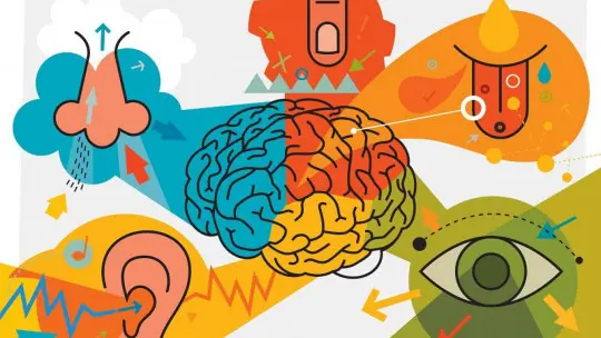
Sight is one of the most evolved and important senses in the human being. Thanks to it we can see the existence of advantageous or threatening stimuli or situations around us with a high level of precision, especially in daylight (for example, it allows us to observe if there are predators in the environment or if we have any type of food available). .
But seeing is not as simple a process as it may seem: it is not only necessary to capture the image but also interpret its parameters, distance, shape, color, and even movement. At the brain level, these processes require processing that takes place in different brain regions. In this sense, highlights the role of the brain’s visual cortex
Throughout this article we will see what the characteristics and parts of the visual cortex are, through a summary about this part of the human brain.
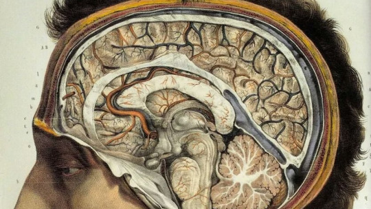
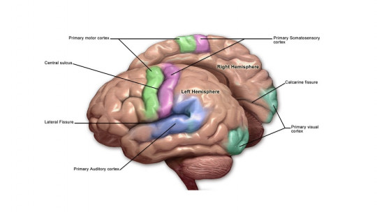
The part of the cortex mainly dedicated to vision is known as the visual cortex. processing of visual stimulation from retinal photoreceptors It is one of the most represented senses at the level of the cortex, its processing occupying most of the occipital lobe and a small part of the parietal lobes.
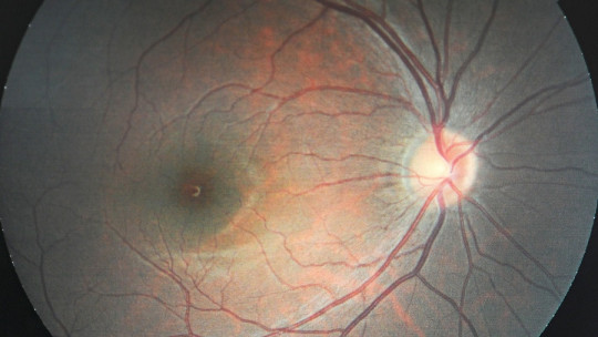
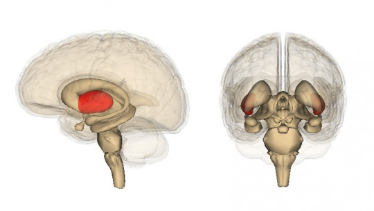
Visual information passes from the eyes to the lateral geniculate nucleus of the thalamus and the superior colliculus, ipsilaterally, to finally reach the cerebral cortex for processing. Once there, the different information captured by the receivers is worked on and integrated to give it meaning and allow us the real perception of fundamental aspects such as distance, color, shape, depth or movement and finally to give them a joint meaning.
However, the total integration of visual information (that is, the last step of its processing) does not take place in the visual cortex, but in networks of neurons distributed throughout the rest of the cerebral cortex.
The visual cortex is not made up of a single uniform structure, but rather includes different brain areas and pathways In this sense, we can find the primary visual cortex (or V1) and the extrastriate cortex, which in turn is subdivided into different areas (V2, V3, V4, V5, V6).
1. Primary visual cortex
The primary visual cortex, also called striate cortex, is the first cortical area that receives visual information and carries out the first processing of it. It is made up of both simple cells (which respond only to stimulations with a specific position in the visual field and analyze very specific fields) and complex cells (which capture broader visual campuses), and is organized in a total of six layers. The most relevant of all of them is number 4, as it is where information from the geniculate nucleus is received.
In addition to the above, it must be taken into account that this cortex is organized into hypercolumns, composed of functional columns of cells that capture similar elements of visual information These columns capture a first impression of the orientation and ocular predominance, depth and movement (what happens in the columns called interblob) or a first impression of the color (in the columns or blob regions also known as spots or drops).
In addition to the above, which the primary visual cortex begins to process on its own, it should be noted that in this brain region there is a retinotopic representation of the eye a topographic map of vision similar to that of Penfield’s homunculus as far as the somatosensory and motor system is concerned.
2. Extrastriate or associative cortex
In addition to the primary visual cortex, we can find various associative brain areas of great importance in the processing of different characteristics and elements of visual information. Technically there are around thirty areas, but the most relevant are those coded from V2 (remember that the primary visual cortex would correspond to V1) to V8. Part of the information obtained in the processing of the secondary areas will later be analyzed again in the primary area to be reanalyzed.
Their functions are diverse and they handle different information. For example, area V2 receives color information from the regions and information regarding spatial orientation and movement from the interblob. The information passes through this area before going to any other, forming part of all visual pathways. Area V3 contains a representation of the lower visual field and has directional selectivity, while the ventral posterior area has the upper visual field determined with color and orientation selectivity.
V4 participates in the processing of stimulus shape information and its recognition. Area V5 (also called medial temporal area) is mainly involved in the detection and processing of stimulus movement and depth, being the main region responsible for the perception of these aspects. The V8 has color perception functions.
To better understand how visual perception works, however, it is advisable to analyze the passage of information through different routes.
Main pathways of visual processing
The processing of visual information is not something static, but rather occurs along different visual pathways of the brain , in which the information is transmitted. In this sense, the ventral and dorsal pathways stand out.
1. Ventral route
The ventral pathway, also known as the “what” pathway, is one of the main visual pathways of the brain, which would go from V1 towards the temporal lobe Areas such as V2 and V4 are part of it, and are mainly responsible for observing the shape and color of objects, as well as depth perception. In short, it allows us to observe what we are observing.
Likewise, it is in this pathway where stimuli can be compared with memories when passing through the lower part of the temporal lobe, such as in areas such as the fusiform in the case of face recognition.
2. Dorsal route
Regarding the dorsal route, it runs through the upper part of the skull, going towards the parietal. It is called the “where” route , since it works especially with aspects such as movement and spatial location. The participation of the V5 visual cortex stands out, with a great role in this type of processing. It allows you to visualize where and how far away the stimulus is, whether it moves or not and its speed.
Alterations caused by injury to the different visual pathways
The visual cortex is an element of great importance for us, but sometimes different injuries can occur that can alter and endanger its functionality.
Damage or disconnection of the primary visual cortex generates what is known as cortical blindness, in which even though the subject’s eyes function correctly and receive the information, it cannot be processed by the brain, so the to perceive. Also Hemianopsia may appear if damage occurs in only one hemisphere blindness appearing only in one visual hemifield
Lesions in other brain regions can cause different visual alterations. A lesion of the ventral pathway will probably generate some type of visual agnosia (either apperceptive in which it is not perceived or associative in which although it is perceived it is not related to emotions, concepts or memories), by not being able to recognize the objects and stimuli that are presented to us. For example, it could generate prosopagnosia or lack of identification of faces at a conscious level (although not necessarily at an emotional level).
Damage to the dorsal pathway could cause akinetopsia inability to detect movement visually.
Another probable alteration is the presence of problems when it comes to having a consistent perception of space, not being able to consciously perceive a part of the visual field. This is what happens in the aforementioned hemianopia or quadrantpsia (in this case we would be facing a problem in one of the quadrants).
Likewise, vision problems may appear such as difficulties with depth perception or blurred vision (similar to what happens with eye problems such as myopia and hyperopia). Problems similar to color blindness (we are talking about monochromatism or dichromatism) or lack of color recognition may also appear.