Inferior Frontal Gyrus Of The Brain: Its Characteristics And Functions
The brain contains numerous folds and gyri that make up different anatomical structures, among which are the lower frontal gyrus, which we will talk about in this article
This brain region is part of the prefrontal cortex, and houses areas as important as Broca’s, essential in language production.
Below we explain what the inferior frontal gyrus is and where it is located, what functions it performs and what are the main disorders associated with injuries to this area of the brain.
Inferior frontal gyrus: definition, structure and anatomical location
The lower front turn is one of the many convolutions contained in the human brain ; folds that make up the characteristic relief of this organ and that give that wrinkled appearance to its outer surface, the cerebral cortex.
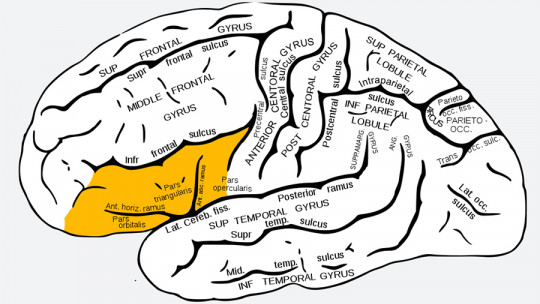
This gyrus is located below the inferior frontal sulcus, and extends forward from the bottom of the precentral sulcus. It is delimited by its anterior part with the lateral or Sylvian fissure. Up to three different parts can be identified in the inferior frontal gyrus: the opercular pair, behind the anterior ascending ramus; the triangular pair, between the ascending and horizontal branches; and the orbital pair, below the anterior horizontal branch of the fissure.
The caudal limit of the pars opercularis is the inferior precentral sulcus and its rostral limit is the anterior ascending branch of the lateral fissure Sometimes, an additional sulcus is usually identified: the diagonal sulcus, within the opercular pairs. When this is present, it may remain separate or may be mixed with the ascending sulcus.
On the other hand, there are authors who have divided the inferior frontal gyrus into a posterior part and an anterior part. While it is true that this sulcus can be said to continue, ventrally, almost to the lateral margin of the orbital frontal region, this may be a false impression as a result of the fusion of the anterior part of the inferior frontal sulcus with a different sulcus. which often forms the anterior end of the triangular pair: the pretriangular sulcus.
Features
The inferior frontal gyrus is one of the parts that make up the prefrontal cortex of the brain, whose main functions have to do with executive control and planning of complex behaviors, decision making or management, and adaptation of behavior to social norms
In recent years, research has focused on the role of the inferior frontal gyrus in a specific aspect of executive control: behavioral inhibition or response inhibition This can be defined, in general terms, as the ability we have to control and retain responses to routine or predominant internal or external stimuli that appear during the performance of a task.
Tasks examining response inhibition typically involve the development of a routine response, followed by its cancellation when an infrequent stop signal is detected. For example, with the Go/No go task, in which there are two starting conditions, and in some trials you have to respond to the stimuli (Go trial) and in others you do not have to respond (No go), so that the The examiner can then measure the individual’s ability to inhibit his or her responses.
Another function in which the inferior frontal gyrus would be involved is attentional control. To measure the attentional capacity of a subject, “Stop signal” tasks are usually used, an experimental test that serves to measure the inhibitory processes and the automaticity of the subjects’ responses, and that also uses Go/Go tasks. Don’t Go.
The latest findings based on studies carried out with functional magnetic resonance imaging confirm that the inferior frontal gyrus also plays a general role in attentional control as a necessary structure for the person to adapt and respond to relevant stimuli and become inhibited in the face of non-relevant or distracting stimuli.
It is also worth noting the role that the inferior frontal gyrus plays in language processing, since Broca’s area is part of this gyrus. This brain region, located in the left hemisphere, is essential in the expression of language and speech production, since it is responsible for planning the sequence of movements necessary for us to articulate the words we pronounce.
mirror neurons
Mirror neurons are a special class of neurons that are activated both when observing the behavior of others and when executing it ourselves. They are called mirrors because they allow us to deduce what others think and feel, and they are closely related to aspects such as empathy, imitation or social behavior.
There is evidence that there are mirror neurons in various brain regions, including: the pars opercularis of the inferior frontal gyrus and the inferior parietal lobe, although it is also suggested that there could be nuclei of these neurons in other areas such as the insula, the anterior cingulate and the superior temporal gyrus.
Studies carried out with people with autism spectrum disorder (ASD) show the existence of alterations in the activity of their mirror neurons, specifically in the inferior frontal gyrus, the insula and the cingulate cortex. These alterations would explain the inability of these people to grasp the intentions of others and experience empathy just as people without ASD do.
Related disorders
Research carried out on patients with lesions in the prefrontal lobe that include the inferior frontal gyrus have revealed that they usually present a deficit in response inhibition. It has been suggested that there is a centrally located inhibitory mechanism that suppresses irrelevant responses, and that this inhibition would be located mainly in the right inferior frontal gyrus.
On the other hand, the same inhibitory control mechanism appears to be altered in patients with obsessive-compulsive disorder (OCD) In a study in which healthy subjects were compared with individuals with this disorder, significant alterations in structural connectivity were found, probably associated with a lack of myelination and axonal problems in the inferior frontal gyrus of patients with OCD.
Finally, it has been proven that lesions in Broca’s area, which corresponds to Brodmann’s area 44 and area 45 (opercular pair and triangular pair of the inferior frontal gyrus in the left hemisphere, respectively) involve linguistic difficulties like the ones presented below: