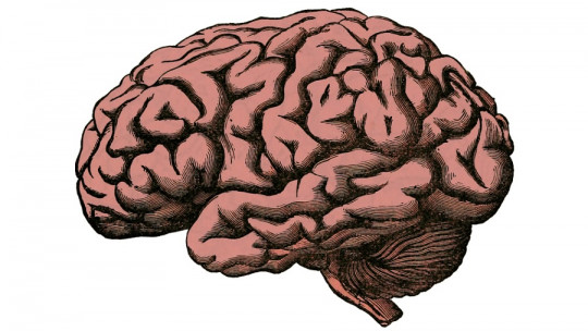
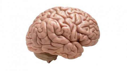
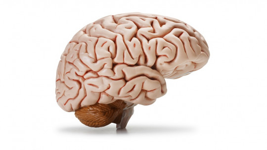
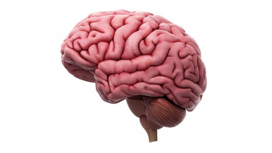
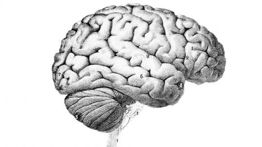
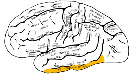
Throughout evolution, the brain has become more complex by optimizing the way it organizes its structure, using a resource as valuable as fissures or folds, small indentations and grooves with which it extends its surface by folding inward.
This mechanism has allowed our species to improve certain higher cognitive functions. We will describe the most relevant fissures, including the gyri and sulci, of our brain.
Brain fissures , also known as sulci, are the prominent grooves that traverse the surface of the cerebral cortex, dividing it into distinct gyri or ridges. These intricate folds are a hallmark feature of the human brain, contributing to its remarkable complexity and functional specialization. In this article, we delve into the anatomy of brain fissures, their developmental origins, and their significance in brain function and evolution.
What are the fissures of the brain?
The human brain is an extremely complex organ Made up of millions of nerve cells, as well as glial cells and blood vessels. It is a fundamental part of the central nervous system, responsible for centralizing and processing information from our body and the environment to generate the best possible responses, depending on what each situation demands.
The brain can be divided into hemispheres: the right hemisphere and the left hemisphere; and in turn, in lobes: the frontal lobe, which is responsible for language and executive functions; the temporal lobe, responsible for hearing or speech; the parietal lobe, responsible for sensory-perceptive functions; the occipital lobe, whose main function is visual processing; and the insula or insular cortex, which separates the temporal and inferior parietal lobes and plays a key role in emotional processing and subjective experience.
In neuroanatomy, when describing the different brain structures, the fissures are taken into account, which cover the surface of the cortex of the brain and They give it that peculiar rough characteristic These “wrinkles” are essential for this organ to function correctly; An absence of these can cause serious disorders, such as lissencephaly (or “smooth brain”), which can cause motor problems, seizures and other alterations.
The fissures of the brain They can be divided into gyri and sulci that are found on the entire surface of the cortex , demarcating the different cerebral lobes and hemispheres, and allowing their extension to be greater; In such a way that, evolutionarily speaking, the more the brain has retracted inward, the greater complexity it has gained over the years, with the consequent increase and improvement of certain cognitive functions in the human species, such as language or intelligence.
Anatomy of Brain Fissures
Sulci and Gyri
Brain fissures are formed by the infolding of the cerebral cortex, resulting in the formation of deep grooves known as sulci and raised ridges called gyri. Sulci vary in depth and complexity, with some extending deeply into the cortical tissue, while others are more shallow and superficial. Gyri, on the other hand, represent the elevated regions of the cortex between sulci and are often associated with specific functional areas of the brain.
Major Sulci of the Brain
Several major sulci are recognized in the human brain, each with its own distinct characteristics and functional significance. Examples include the central sulcus, which separates the frontal and parietal lobes and plays a critical role in motor and sensory processing, and the lateral sulcus, which demarcates the temporal lobe and houses important structures involved in auditory processing and language comprehension.
Developmental Origins
Genetic and Environmental Factors
The formation of brain fissures is a complex and tightly regulated process influenced by both genetic and environmental factors. Genetic mechanisms govern the proliferation, migration, and differentiation of neural progenitor cells during embryonic development, shaping the overall architecture of the cerebral cortex. Environmental factors, such as intrauterine conditions and maternal health, can also impact cortical development and contribute to variations in sulcal patterns among individuals.
Role of Mechanical Forces
Emerging evidence suggests that mechanical forces play a significant role in shaping brain fissures during fetal development. As the brain undergoes rapid growth and expansion within the confined space of the skull, mechanical tension and compression exerted by surrounding tissues and cerebrospinal fluid contribute to the formation of sulci and gyri. This process, known as gyrification, results in the characteristic convoluted appearance of the mature brain and is thought to enhance cortical surface area and neuronal connectivity.
Functional Significance
Increased Surface Area
The folding of the cerebral cortex into sulci and gyri significantly increases its surface area, allowing for greater neuronal density and connectivity within a compact spatial footprint. This structural complexity is thought to facilitate efficient information processing, enabling the brain to integrate sensory inputs, coordinate motor functions, and support higher-order cognitive processes such as memory, attention, and language.
Functional Localization
Brain fissures play a crucial role in functional localization, with specific regions of the cortex associated with different sensory, motor, and cognitive functions. The topographic organization of sulci and gyri reflects the specialized functions of underlying cortical areas, allowing for precise mapping of brain activity using neuroimaging techniques such as functional magnetic resonance imaging (fMRI) and electroencephalography (EEG). By delineating distinct functional domains, brain fissures aid in the identification and study of neural circuits underlying behavior and cognition.
Features and functions
The fissures of the brain, whether they are gyri or grooves of greater or lesser depth, fulfill important functions; On the one hand, as we mentioned in the introduction, These folds increase the surface of the cerebral cortex and neuronal density (without having to increase the size of the head), with the consequent improvement of higher cognitive functions in the medium and long term.
At an evolutionary level, this represents a great qualitative leap, since otherwise, increasing the size of the head and skull would only have been a problem for childbirth in women.
According to most scientific studies, this folding most frequently occurs in species with larger brains, like ours, although there seem to be exceptions (as is the case of manatees, with fewer folds than expected for a brain of its size).
However, the formation of fissures depends on other factors beyond the growth and expansion of the surface of the cerebral cortex, such as the physical properties of some parts of the cerebral cortex; For example, thinner regions of the brain tend to bend more easily and the brain folds into specific, consistent patterns
On the other hand, although the brain is an interconnected organ, the different fissures are used to separate and delimit areas and structures with different functions, acting as borders that help in the division of tasks.
The main grooves of the brain
There are many grooves or grooves in the brain. Next, we will talk about the most well-known and relevant ones.
1. The interhemispheric groove
The interhemispheric sulcus or fissure, also known as the longitudinal fissure, is a groove located in the cortex that divides the brain into two hemispheres, joined together by a set of nerve fibers called the corpus callosum. This fissure contains a fold of the dura mater (the outer meninge that protects the central nervous system) and the anterior cerebral artery
2. The lateral groove
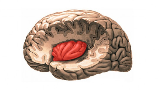
The lateral sulcus or Sylvian fissure is one of the most visible in the brain, since it runs transversely across practically the entire surface of its cortex. It is located in the lower part of the brain hemispheres , delimiting the border between the temporal lobe and the parietal lobe. It is also one of the deepest crevices, and beneath it is another relevant structure of the brain: the insula.
3. The central groove
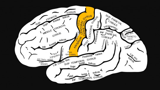
The central sulcus or Rolando fissure is a groove located in the upper part of the brain and separates the frontal lobe from the temporal lobe, bordering on one side with the motor cortex and, on the other side, with the primary somatosensory cortex. This fissure would act as a bridge between motor and sensory information, integrating both.
4. The parieto-occipital sulcus
The parietocipital sulcus or external perpendicular fissure It is a cleft that originates in the interhemispheric fissure , being present on the inner side of each cerebral hemisphere. As its name suggests, it separates the parietal lobe from the occipital lobe.
The lateral part of the sulcus is located in front of the occipital pole of the brain and the medial part runs downwards and forwards. It joins the calcarine fissure below and behind the posterior end of the corpus callosum.
5. The calcarine groove
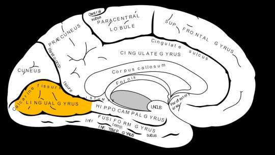
The calcarine sulcus or fissure is a groove located in the occipital area of the internal or medial side of the cerebral hemispheres, separating the visual cortex into two parts. It follows a horizontal path until it joins the parieto-occipital sulcus
6. The callous groove
The callosal sulcus is located on the medial brain surface and separates the corpus callosum from the cingulate, which performs relevant functions within the limbic system. Although the cingulate is usually defined as a separate structure, it is part of the frontal and parietal lobes.
The main convolutions of the brain
As with the sulci that we have seen previously, in the brain there are also a multitude of fissures in the form of convolutions or gyri, characterized by being folds with less depth than the grooves and located inside the different brain lobes. Next, we will see some of the most important ones.
1. Fusiform gyrus or gyrus
The fusiform gyrus or gyrus is located on the basal surface of the cerebral hemisphere, specifically in the temporal lobe, between the inferior temporal gyrus (outside) and the hippocampal gyrus (inside).
This fissure is part of the limbic system , responsible for affective processing and has an important role in facial recognition; Damage to this area of the brain can cause prosopagnosia, also called face blindness.
2. Convolution or cingulate gyrus
The cingulate gyrus or gyrus is an arc-shaped fissure or fold of the brain, located above the corpus callosum. Its main function is act as a link or bridge between the limbic system and higher cognitive functions located in the neocortex so it has a fundamental role in connecting volitional, motor, mnesic, cognitive and affective aspects.
3. Convolution or angular gyrus
The gyrus or angular gyrus is a fissure located in the parietal lobe, more specifically between the intraparietal sulcus and the horizontal branch of the Sylvian fissure.
The functions of the angular gyrus include processing and interpreting language, visual and auditory information It has connections with Wernicke’s area, responsible for the auditory decoding of linguistic information.
4. Hippocampal gyrus or gyrus
This gyrus is located in the inner part of the temporal lobe, surrounding the hippocampus, a fundamental structure in the formation of new memories and spatial location.
Clinical Implications
Neurodevelopmental Disorders
Abnormalities in brain fissures have been implicated in various neurodevelopmental disorders, including autism spectrum disorder, schizophrenia, and epilepsy. Disruptions in cortical folding patterns may reflect underlying disturbances in neurogenesis, neuronal migration, or synaptic connectivity during early brain development, leading to altered brain structure and function. Characterizing sulcal abnormalities using neuroimaging techniques can aid in the early diagnosis and intervention of neurodevelopmental disorders.
Neurosurgical Planning
Brain fissures serve as important landmarks for neurosurgical planning and navigation, particularly in procedures involving the removal of brain tumors or the treatment of epilepsy. Surgeons rely on preoperative imaging studies to identify the location and extent of sulci and gyri, allowing for precise targeting of surgical incisions while minimizing damage to adjacent functional areas. Advances in neuroimaging technology, such as diffusion tensor imaging (DTI) and tractography, have further improved surgical outcomes by providing detailed maps of white matter tracts and cortical connectivity.
Brain fissures represent a striking example of the intricate interplay between genetic programming, mechanical forces, and environmental influences during brain development. Their convoluted patterns reflect the remarkable complexity and functional specialization of the human cortex, supporting a diverse array of cognitive, sensory, and motor functions. By unraveling the anatomy, developmental origins, and functional significance of brain fissures, researchers and clinicians gain valuable insights into the organization and operation of the human brain, paving the way for advances in neuroscience, neurology, and neurosurgery.

