The enteric nervous system is the part of the autonomic nervous system responsible for regulating vital gastrointestinal functions. Among these functions is the regulation of the esophagus, stomach and colorectal functions; which in turn involves the absorption and digestion of nutrients, as well as the maintenance of protective mucous membranes. The functioning of this system is the most complex of the set of elements that make up the autonomic nervous system.
Below we will see in more detail what the enteric nervous system is and what some of its main functions and characteristics are.
The enteric nervous system is the cellular structure responsible for controlling our gastrointestinal functions. The above includes the mobility, secretion, local immunity and inflammation of the organs that make up the digestive system
In other words, the enteric nervous system is responsible for regulating important functions for the intake, absorption, metabolism and digestion of food. It is also responsible for preventing diseases related to these activities.
The enteric nervous system originates in the cells of the neural crest (a structure generated during embryonic development), which, in turn, is divided into two large branches of intertwined nerve cells. These branches are called “Meissner’s submucosal” and “Auerbach’s myenteric” branches, and they make up the two main components of the enteric nervous system.
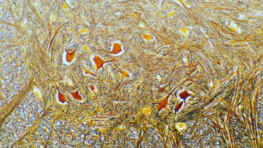
This system is recognized as the most complex part of the peripheral nervous system and It is composed of a high concentration of neurons and glial cells In fact, it contains the largest collection of neurons found outside the brain.
Origins and development of this system
The enteric nervous system is formed from embryonic development, from two main processes: cell proliferation and its differentiation with the great variety of glial cells and neuronal variants that make up the organism.
From the fourth week of gestation, a part of the neural crest cells, which give rise to the majority of the enteric nervous system, migrate through the entire digestive tract
The other part of the same cells, which contributes to a lesser extent to the formation of the ENS, migrate from the cranial region to the caudal region (i.e., from the head to the opposite end). The latter gradually spread through the gastrointestinal tract of the embryo in all its components:
Components of the SNE
As we saw before, the enteric nervous system is divided into two main segments that arise from the neural crest. Each of them contains a wide variety of glial and nerve cells, and they are jointly responsible for regulating the intake, absorption and metabolism of everything we ingest. These segments, according to Oswaldo, et al. (2012), are the following:
Meissner’s submucosal plexus
It develops mainly in the small intestine and colon, and It is responsible for regulating digestion and absorption in music and blood vessels
Auerbach’s myenteric plexus
It is found throughout the digestive tract, and is responsible for coordinate the activity of the muscular layers of said organ
The large number of enteric neurons in the healthy adult small intestine remains constant throughout most of adult life, which appears to be the result of a process of continuous renewal of neurons in the intestine (Kulkarni, S. et al, 2017). .
The neurons that are part of the enteric nervous system, and therefore are responsible for regulating our gastrointestinal activity, are the following (Oswaldo, et al, 2012):
1. Primary intrinsic afferent neurons
Being afferent, these are neurons that transport nerve impulses from the organs to the central nervous system. However, being primary neurons, they do not conduct sensory information directly, but do so through other cells located in the enteric epithelium (the cellular tissue that lines the enteric nervous system). That is to say, Its activity is mainly that of sensory transducers and in this way they regulate physiological functions of the digestive tract.
2. Motor neurons
As its name says, it is responsible for activating the muscular layers that make up both the digestive tract and the blood vessels and some glands. They are further divided into excitatory motor neurons (for example, acetylcholine), or inhibitory motor neurons (such as nitric oxide or GABA). The latter, inhibitory neurons, are responsible for regulating water secretion, blood flow and the release of electrolytes.
3. Interneurons
These are the nerve cells responsible for connecting primary intrinsic afferent neurons with motor neurons. They can be ascending or descending depending on whether they act from the head to the opposite end, or in the opposite direction.
Its extensions are located outside the digestive tube and connects with the nervous ganglia to form a new ganglion called “prevertebral”. Its main function is to warn about changes in the activity of the intestine, so it is mechanoreceptors (secondary neurons that fire action potentials upon mechanical stimuli).
Main functions of the SNE and associated pathologies
According to Furness, 2012, the main functions performed by the enteric nervous system as a whole are the following:
Improper operation of this system affects the functions described above. For the most part, the inadequate functioning of the SNE It is related to neuropathies that make it difficult to control muscle activity and the movement of mucosal fluid The above is reflected in different conditions of the colon and digestive tract.
Furthermore, inadequate functioning of the ENS may be of congenital origin or acquired during postnatal development. Generally, the latter occurs due to a secondary medical condition that ends up significantly damaging the functioning of the ENS, although it can also occur due to an iatrogenic effect of a drug, or due to neuropathology induced by drug use.
: Parts and Functions")


