The 10 Parts Of The Ear And The Sound Reception Process
The auditory system is relatively simple compared to those of the other senses; This is because the process by which sound vibrations are converted into nerve impulses It has a linear character. Sound is transmitted from the ear to the auditory nerve, and from this to the brain, by a chain of internal structures.
In this article we will describe the outer, middle and inner ear, the main components of the auditory system, as well as the substructures that form each of these sections. To complete this description we will explain the process by which air vibrations become sounds perceptible to humans.
Parts of the outer ear: from the ear to the eardrum
The outer ear It is made up of the ear, the ear canal and the eardrum or tympanic membrane. The function of this segment of the auditory system is to capture sound vibrations and channel them to the innermost parts of the ear. In this process, some of the collected frequencies are increased and others reduced, so that the sound is modified.
1. Ear or pinna
The ear is the most external component of the auditory system, and the only one that can be seen from the outside. This structure, also known as the “auricle,” is made up of cartilage and skin. Its function is to collect auditory energy and redirect it to the middle ear through the ear canal.
The ear canal is a cavity that connects the ear to the eardrum. Sound vibrations reach the middle ear through this canal, which measures approximately 2.5 to 3 centimeters long and just 7 square millimeters in diameter.
3. Eardrum or tympanic membrane
The eardrum is a membrane that separates the outer ear and the middle ear ; Strictly speaking, it is not part of any of these segments, but rather it is the structure used to delimit them. It is also known as the “tympanic membrane”.
Middle ear: the chain of ossicles
After reaching the eardrum, the sound vibrations are transmitted through the ossicles of the middle ear to the oval window of the cochlea, where the transduction into nerve impulses will take place.
1. Hammer, anvil and stirrup
The ossicular chain is made up of the hammer, incus, and stapes Amphibians, reptiles and birds have only one bone, the columella, which is morphologically equivalent to the stapes of mammals.
The malleus is attached to the eardrum, while the stapes connects to the cochlea; The transmission of vibrations through the ossicles causes the lymph fluid in the inner ear to move, a necessary step for sound transduction.
2. Oval window
The oval window is the membrane that covers the cochlea, so it is technically located between the inner and middle ear. The vibrations in the eardrum are transmitted through the ossicles to the oval window, which consequently also vibrates, stimulating the inner ear.
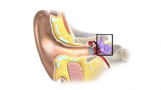
Inner ear: the cochlea and transduction
The inner ear is a cavity located inside the skull. It is here where the transduction of sound vibrations into nerve impulses takes place, which marks the beginning of brain processing of hearing.
The key structure of the inner ear is the cochlea or cochlea, a set of channels that rotate on themselves and that amplify the auditory signals they receive. Inside the cochlea is the organ of Corti, mainly responsible for hearing.
1. Semicircular canals
The semicircular canals or ducts are an organ of the inner ear composed of two compartments, the saccule and the utricle, which allow the sense of balance in association with the ossicular chain.
2. Vestibular scale or higher
The oval window of the cochlea, which is located in the scala vestibular, connects the stapes with the rest of the inner ear. This structure is full of perilymph a substance similar to cerebrospinal fluid that receives vibrations from the ossicular chain.
3. Scala tympani or inferior
The sound waves received by the upper scala are transmitted to the lower scala through the perilymph since the two structures are connected by this fluid, while the basilar membrane separates them.
4. Cochlear or average scale
The cochlear scala is isolated from the vestibular and tympanic membranes by the Reissner membrane and the basilar membrane, respectively; however, it also shares endolymph with other parts of the inner ear.
The organ of Corti is located in the middle scale, where the transduction of sound vibrations into neural impulses takes place. The hair cells found in this structure allow transduction.
5. Auditory or vestibulocochlear nerve
The vestibulocochlear or auditory nerve, composed of the cochlear and vestibular nerves, transmits information about sound and balance. from the inner ear to the central nervous system The vestibulocochlear nerves constitute the eighth of the twelve cranial nerves.


")
