The musculoskeletal system refers to the set of organs and structures that allow us to move in three-dimensional space and maintain posture despite the gravitational force. Without it, we would surely be like a worm or a small nemertine, stuck to the ground and making slow and costly movements in the horizontal plane, with a flattened body and basic morphology. Can you imagine what human life would be like without muscles and a skeleton?
The musculoskeletal system includes the osteoarticular system (bones, joints and ligaments) and the muscular system (muscles and tendons). This true work of art of biomechanics allows us to interact with the environment and in turn support the different organs of the body without collapsing. Something as simple as getting out of bed would be impossible without the bones and muscles involved.
Today we went down in scale drastically. We have already covered the skeletal system, isolated parts of the skeleton, the human musculature, the facial musculature and many other thematic fronts more associated with the musculoskeletal system. In this case, we approach a tissue level, much more basic, but just as important as the most complex system of living beings: stay with us if you want to know everything about muscle fiber
Muscle fibers, as their name suggests, make up muscles. Therefore, to understand them we must take a short trip through the muscular system in general and the types of muscles that can be observed. We don’t delay.
The muscular system refers, in general, to all the muscles that can be contracted voluntarily by the body Other authors argue that the heart muscles or those that promote peristaltic movements in the intestines should also be included in this group, but these are usually left out, since their action is independent of individual desire.
If we count only the muscles associated with bones that respond voluntarily to brain commands, we would say that the muscular system is made up of about 650 muscular units. If we also take into account the involuntary muscles, this figure would easily increase above 800. Be that as it may, in our body there are 3 types of muscles:
Approximately 40% of the weight of an adult human being corresponds to skeletal muscle tissue On the other hand, only 10% (at most) is smooth muscle. There are many more skeletal muscles than smooth muscles, but they are all essential to maintaining the individual over time.
After these lines, we get a slight idea of what the muscular apparatus is and what types of muscles make it up (or are left out). Now we are ready to dissect the muscle fiber in its entirety.
What is a muscle fiber?
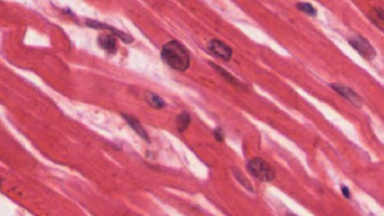
The muscle fiber (or skeletal myocyte) is a multinucleated cell or syncytium. This last term refers to a cell body that has several nuclei, due to the fusion of several cells. Since most cells in eukaryotic multicellular organisms have a single nucleus and a well-defined cytoplasm, the syncytium is a special structure worth mentioning.
Continuing with the classic definition, we can say that A muscle fiber is the cell type that makes up the skeletal or striated muscle tissue, that is, the one that is attached to the bones and causes conscious movements in the human being The main characteristic of this cell body will, therefore, be contractility: the ability to shorten its own length, triggering work in doing so.
From here, things get a little complex. It is best to imagine the cross section of a muscle as a large cable in which many other small cables have been stored. We explain ourselves in the following lines.
The organization of muscle fibers
If you perform a cross section of a circular muscle, the first thing you will find in the outermost part is the epimysium, a layer of connective tissue that is in direct contact with the external environment. If you look closely, you will see that within the large circle that is the cross section, there are other smaller circles grouped together. These are the fascicles, which are surrounded by another layer, known as perimysium.
Within the fascicle we find the muscle fibers themselves, arranged in a bundle Reviewing what we have learned so far:
Muscle section (epymysium) > various fascicles (perymysium) > Muscle fibers
Making an analogy, it is as if various smaller but also large cables (fascicles) were introduced into the sheath of a large diameter cable (muscle), and within these is where the conductive elements (muscle fibers) would actually be. Has this become a little clearer?
Muscle fiber anatomy
The complexity did not end there, since we have described where the muscle fiber is located, but not what it is composed of. As a cell, it must have organelles, cytoplasm and nucleus, TRUE? That’s right, but in this case, the myofibrils occupy a large part of the cellular space, completely changing the typical arrangement of their structures.
We start with the basics: the muscle fiber has a plasma membrane, like the rest of the cells of living beings. It is a semipermeable and lipid membrane, however, it extends in the form of trabeculae inside the cell. This membrane is known as the sarcolemma
Like any other cell, the muscle fiber also needs a cytoplasm in which the rest of the substances are housed, and in this case, It is known as sarcoplasma This is composed of a solution phase based on water, ions and small diffusible molecules, which surrounds fixed macromolecular structures, the myofibrils.
Like every cellular body, muscle fibers also need energy. Therefore, mitochondria appear between the myofibrils, tightly packed and in contact with each other. The mitochondria are located practically attached to the myofibrils, as they need to provide all the energy necessary for the contraction process, which is not exactly small. The sarcoplasmic reticulum also surrounds the myofibrils, as it stores the calcium necessary to begin the cascade reaction of muscle contraction.
The sarcoplasm (remember that it is the analogue of the cytoplasm) of a muscle fiber has a huge number of myofibrils inside: we are talking about several hundred or even thousands of them. Each myofibril alone contains about 1,500 myosin filaments and 3,000 actin filaments. These biopolymers are responsible for the contraction of the myofibril, and therefore of the muscle fiber, until the entire muscle is reached.
Finally, it is essential to highlight that This cell type is part of a stable tissue with very little nuclei rotation For this reason, the turnover rate of muscle fibers does not exceed 1-2% per week, a very low figure when compared to the turnover rates of the most superficial layer of the epidermis, for example.
There are slow-twitch and fast-twitch fibers, which will determine the functionality and effectiveness of the muscle tissue depending on the task to be performed. We will explore this physiological diversity in future occasions.
Summary
What do you think? It is very curious to know that, at a microscopic level, some of the cells in our body have gone through drastic change processes in order to acquire specialized functionality. The muscle fiber is a clear example of this: It is the product of several cells, it has several nuclei, it is separated from the medium by a sarcolemma and within its sarcoplasm it houses thousands of myofibrils so that its contraction can occur.
Thanks to these physiological specializations, many cells are capable of performing highly specialized tasks inconceivable without them. Without the muscle fiber, the movement and permanence of the human being as we know it today in the three-dimensional environment would be completely impossible.

")


