Baciloscopy: What It Is And How It Is Used In Medicine
Baciloscopy is a test performed in medicine to detect bacillus-type bacteria in a given sample. This methodology is very useful in the identification of the microorganism Mycobacterium tuberculosis, the causative agent of tuberculosis (TB).
In the world of microbiology, detection is the key to success. In order to treat an infectious disease, finding the causative agent quickly before it multiplies uncontrollably is essential. This can be simple in parasitic processes such as taeniasis, for example, where the parasite measures more than one meter.
Unfortunately, bacteria are much smaller and elusive in many cases. For this reason, sophisticated methods have been devised for its detection, such as the smear microscopy that concerns us today. If you want to know more about the topic, we encourage you to continue reading.
Since we cannot start building the house from the roof, we must first understand what a bacillus is, so that later we can delve into how to detect it.
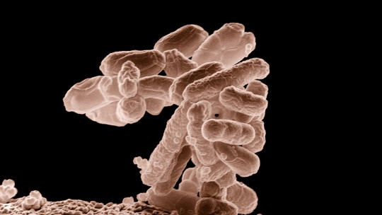
The word “bacillus” is used to describe any rod- or rod-shaped bacteria. Thus, it is a morphological classification that does not include species, genera and orders In any case, there is a group that uses this term, bacteria within the genus Bacillus.
It may seem confusing, but this terminological complex can be summarized as follows: all bacteria of the genus Bacillus They are bacilli, but not all bacilli belong to this genus Without going any further, the bacteria that causes tuberculosis belongs to the genus Mycobacterium, even though it is rod-shaped. This is not the only one, since among many others, the genera Salmonella, Moraxella or Yersinia are also considered bacilli due to their elongated morphology.
We have defined the first key term to understand smear microscopy: the bacillus. This is the causal principle of the test, but the purpose of course is summarized in detecting tuberculosis Therefore, this disease requires special prior mention.
The bacillus of death and tuberculosis
The World Health Organization (WHO) gives us certain relevant data regarding tuberculosis. Some of them are the following:
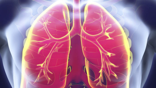
As we can see, we are faced with a pathogenic bacteria extremely harmful to human society The growth of Mycobacterium tuberculosis in the patient’s airways causes a productive cough, chest pains, weakness, weight loss, and night sweats. Through the blood or lymphatic route, bacteria can spread to other organs, aggravating the clinical picture even more.
One of the biggest problems with tuberculosis is that The most obvious symptoms begin to appear when the lesions in the lung tissue are already severe and the infection is in an advanced state Therefore, tests such as smear microscopy are essential to act as quickly and efficiently as possible. Next, we explain what this detection method consists of.
The differential diagnosis
We already know the principle (the bacillus Mycobacterium tuberculosis) and the end (TB tuberculosis). Now, naturally, we are left to immerse ourselves in the world of diagnoses that correlate the microorganism with the patient’s disease. To do this, it is necessary to follow a series of steps.
1. Sample collection
First of all, it is necessary to emphasize that in order to perform the smear microscopy a sputum sample is required from the patient According to clinical studies, it should be stored in a standard container (wide-mouth, airtight, and made from break-resistant plastic).
As the elimination of bacilli with sputum is not constant, it is recommended that a total of three samples be collected per patient. The first detects approximately 80% of positive cases, the second 15% and the third the remaining 5%. Of course, Samples should be obtained at different times of the day to maximize the possibility of detecting the pathogen
In the case of suspected spread of the infection to other organs, samples of cerebrospinal fluid, urine or pus from an abscess can be taken.
Once obtained, transported and fixed on a slide, it is time to search the sample for the microorganism causing the disease.
2. Staining
In order to observe the bacillus in the sample, it is necessary to subject it to a staining process specifically to the Ziehl-Neelsen stain.
The basis of this technique is based on the fact that the cell wall of certain bacteria (such as Mycobacterium tuberculosis) has acid-alcohol resistant capabilities, that is, it These bacteria have a property of retaining basic dyes despite exposure to decolorizers such as the acid-alcohol complex
For this reason, a dye called fuchsin is applied to the extended sputum sample, which will then be subjected to a discoloration process. After that, a new dye will be used.
Bacteria that present a red color after the discoloration process (due to the fuchsin retained in their cell wall) are those sought, while the rest are observed with a blue color (since methylene blue is used as a subsequent contrast dye). .
Thus, under the microscope you can observe a series of small curved elements, isolated or aggregated, in the shape of a fuchsia-red rod between one and 10 micrometers long. This allows a clear differential diagnosis: If there is a density of red microorganisms among the blue ones in the patient’s sample, tuberculosis is guaranteed
All that glitters is not gold, because despite the speed and cheap cost of smear microscopy, the World Health Organization warns us that this It only detects half of tuberculosis cases and is unable to report whether there is drug resistance on the part of the microorganism
Of course, observing whether or not the bacteria is present in the patient’s sample is the first step, but also understanding whether it is a strain resistant to drugs such as rifampin (multidrug-resistant tuberculosis) can mean the difference between life and death of the patient.
Therefore, this organization recommends performing the “Xpert MTB/RIF” test , which detects the disease and rifampicin resistance simultaneously in less than two hours. Although the sensitivity of this new test is very high against lung infections, its diagnostic capacity decreases when the infection spreads to other organs.
Conclusions
As we have seen, smear microscopy is a method of detecting bacillus-shaped bacteria such as Mycobacterium tuberculosisthe pathogen that causes the infectious disease that causes the most deaths worldwide.
Still, due to the emergence of drug-resistant bacterial strains, it is necessary to further refine detection methods : It is no longer enough to know that the bacteria is present in the patient’s sample, but also to which drugs it responds and to which it is resistant.
For all these reasons, This form of detection is considered relatively archaic and increasingly out of use although it is always a good option in hospitals in low-income countries without sophisticated means.