Neck Muscles: Types, Location, Characteristics And Functions
The human musculoskeletal system is a true work of art at a biomechanical level, as muscles, bones and joints work together with the aim of obtaining the greatest range of movement and functionality with the minimum possible cost. Our osteoarticular system is made up of 206 bones, 360 joints and more than 600 skeletal muscles, which allow voluntary movement of the limbs and head.
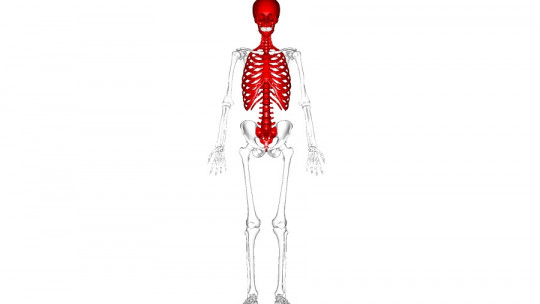
The skeleton, for its part, is made up of two different sections: the axial and the appendicular. The axial skeletal system consists of 80 bones that define the human central axis, that is, the skull, auditory bones, hyoid, rib cage, sternum and vertebral column. On the other hand, the appendicular skeleton is made up of 26 different bones, which define the anatomy and functionality of our limbs.
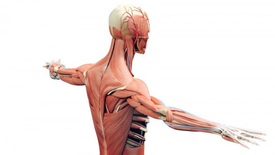
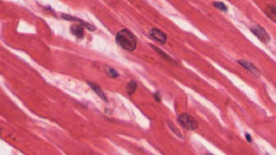
Beyond the skeletal system, the muscular system reports unusual variability. Normally, within the locomotor segment, we only include striated muscles: those that are controlled voluntarily. For this reason, in this topic we leave out the visceral and cardiac muscles, for example, since these move without the human being being completely aware of it.
The face and neck are one of the most complex muscular subsystems of the human being, as they allow us from communication to swallowing and breathing, that is, everything that defines us both as individuals and as a species. In honor of this intricate system, today we tell you about it all about the neck muscles and their particularities
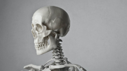
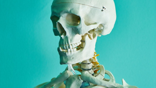
On a physiological level, the neck is nothing more than the junction between the head and the trunk. This structure is home to the proximal esophagus, trachea, thyroid gland, and parathyroid glands in addition to serving as a nervous and blood highway so that all the necessary nutrients and information reach the brain.
It is an integral part of the human organism and stands out for its complexity, since it has many different planes and compartments.
The functionality of the neck can be divided into different fronts and facets These are the following:
As you can see, The neck is essential for the survival of the human being, at both a physiological, hormonal and anatomical level Without this connecting bridge, it would be impossible to communicate the center of nervous organization (the brain) with the rest of the body (trunk and extremities).
The most important neck muscles
The structures of the neck are divided into three different and well-differentiated planes: the vertebral, visceral and vascular compartments. At the muscular level, three well-differentiated functional groups can be detected Let’s see what they are.
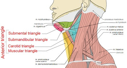
1. Anterior triangle
This section is defined by the anterior border of the sternocleidomastoid muscle, the lower limit of the mandible, and the midline of the neck itself. As its name indicates, These muscle groups cover the anterior face of the neck, that is, what is detected when looking “head-on” at a human being In turn, the previous triangle is divided into various subsections.
1.1 Superficial muscles
They are the ones that are found most externally. Among them, the sternocleidomastoid muscle stands out, located from the sternal manubrium and the medial third of the clavicle to the mastoid process and the superior nuchal line Its functions include contralateral head rotation, ipsilateral tilt, and neck flexion.
1.2 Suprahyoid muscles
As its name indicates, this muscle group is located above the hyoid bone and connects it to the skull. As a group, They support the hyoid and form the muscle mass of the floor of the mouth, allowing us to speak, swallow and breathe among many other things.
This group includes the following components: sternohyoid muscle, geniusohyoid muscle, mylohyoid muscle, and the anterior and posterior portions of the digastric muscle.
The other side of the coin, that is, the muscle sections located below the hyoid bone. They are 4 pairs of muscles in the form of powerful bands located in the front part of the neck which respond to the following designations: the sternocleidohyoid muscle, sternothyroid muscle, thyrohyoid muscle and omohyoid muscle.
As we have said, these are not just one muscle, but we have two of each, one in each “half” of the sagittal plane of the neck. These muscles have different origins and insertions, but their function is unique: to mobilize the hyoid in various directions.
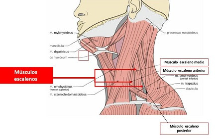
2. Lateral muscles
The lateral muscles are easily palpable, so we encourage you to do the test for yourself when locating them. They are composed of three pairs of muscles: the anterior, middle and posterior scalenus They originate in the CII to CVII cervical vertebrae, and find their insertion point in the 1st and 2nd ribs.
As you can imagine, the main function of these muscle pairs is to elevate the first rib and, in addition, they allow us to tilt our head to the same side: if you tilt your neck to the left side, you will be able to feel the tension of the scalenes on the side. right (and vice versa). They are also essential in the inspiration process.
3. Back triangle
The posterior section of the neck muscles It is composed of those muscle groups that connect the skull with the spine and shoulder girdle (scapulae and clavicles). Again, we differentiate several subgroups within this conglomerate.
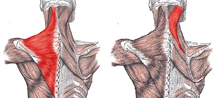
3.1 Surface layer
This includes the trapezius and the splenium (capitus and cervicis). The most important of all is the trapezius, as it connects practically the entire neck to the back Its main functions are the elevation and rotation of the scapula, the stabilization of the scapula, the production of depressing movements and it acts as an extensor of the head and the cervico-dorsal spine.
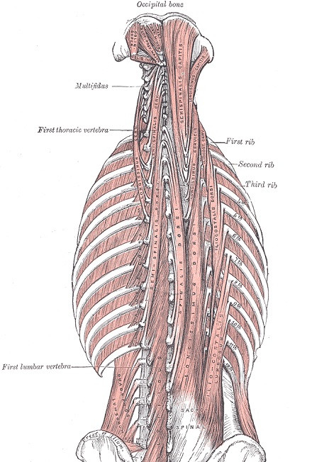
The deep musculature of the posterior neck includes the transversospinalis or transversospinous muscles. Among other things, They are responsible for cervico-cranial stability but we are not going to dwell too much on their particularities due to the anatomical complexity they contain.
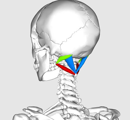
3.3 Deepest layer
Also known as the suboccipital triangle, this layer is composed of the following three muscles: rectus capitis posterior major muscle (superior and medial), superior oblique capitis muscle (superior and lateral), and inferior oblique capitis muscle (inferior and side).
The vertebral artery, the posterior arch of the Atlas (the first cervical vertebra, C1) and the suboccipital nerve pass through this muscular conglomerate. Its clearest function is to provide fine motor function to the head and neck
Summary
As you can see, the neck muscles are divided into seven total sections: three from the anterior triangle, one lateral (on each side of the neck), and three from the posterior triangle. Its functionalities are very varied, as these range from swallowing to structural fixation, including speech, breathing, movement production and many other things.
The head is so versatile at a postural level thanks to these muscle groups, so without them, something as simple as turning the head to avoid danger would be impossible As we gather almost all of our sensitive centers in the head region, it is essential that it be able to adopt different postures and movements, in order to receive the maximum possible information to respond appropriately to environmental pressures.