Osteotendinous Reflexes: What They Are, How They Work, And Associated Pathologies
In neuroscience, a reflex is known as the nervous activity developed in the spine (and brain stem) that consists of an involuntary response to a sensory stimulus, whether internal or external. Generally, we associate reflexes with rapid, uncontrollable jerky movements, but another example of this activity is also the activation of a gland and the secretion of a given compound into the bloodstream.
In any case, at a general level all reflection is involuntary, unplanned, sequential and practically instantaneous. The initiation of a reflex is achieved thanks to the neuronal routes and the reflex arcs, that is, the nervous pathway that runs through the vertebral arch and controls a given reflex act. It should be noted, at this point, that there are 2 types of reflex arcs: the autonomic ones (which affect internal organs) and the somatic ones (which affect the muscles).
With all this information, we are able to paint a general picture that allows us to understand what reflections are and what they are for. In any case, this time we are going to talk specifically about deep tendon reflexes muscle contractions in response to stretching within a muscle.
In humans, when a muscle is hit forcefully, it immediately contracts due to a reflex arc composed of 2 neurons, which also involve the segment of the spinal trunk that innervates the muscular structure analyzed. These are the deep tendon reflexes themselves. For this very special type of reflex to occur, the following physiological elements must be present:
The osteotendinous reflexes that are usually explored, depending on the area stimulated, are the bicipital, tricipital, stylo-radial, ulnopronator, patellar and Achilles reflexes The type of reflex and the response shown always reveal something about the state of the elements of the nervous system involved in its appearance.
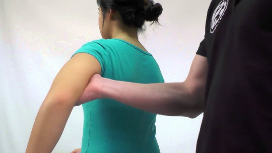
When you want to evaluate the state of the reflex arcs, the professional applies a slight force to an area of the body, which results in a slight elongation of the muscle fiber. This act activates the neuromuscular spindle, formed by a set of sensory receptors inside the muscle that detect changes in its total length.
These receptors They send an afferent impulse to the spinal cord, where a direct synapse occurs with the motor neuron The latter emits the efferent signal back to the muscle, allowing it to contract. As you can see, we are faced with a very simple circuit: it has to be like this, because thanks to the proximity of the structures involved, the deep tendon reflexes occur so quickly.
The medical importance of deep tendon reflexes in Medicine
At this point, it should be noted that there are various conditions that can be suspected based on the patient’s deep tendon reflexes. On the one hand, Hyperreflexia refers to a pathological situation in which the individual suffers hyperactive or repeated reflexes over time (clonics).
Apart from muscle spasms, autonomic hyperreflexia causes changes in heart rate, excessive sweating, high blood pressure and changes in skin color. The most common cause of this clinical entity is a spinal cord injury, although it can also occur due to certain syndromes, side effects to medications, or after severe head trauma.
On the other hand, hyporeflexia and areflexia are events in which the muscle does not produce any response to the application of force It is a situation that reflects a failure or interruption in the reflex arc, either in the efferent or afferent nerve fiber or, on the other hand, it shows conditions in the patient such as hypothyroidism, blood electrolyte alterations or myopathies.
The deep tendon reflex scale
Osteotendinous reflexes They are quantified in the clinic when a nervous or neuromuscular pathology is suspected in the patient To carry out this type of test, the muscular structure to be analyzed must be in a neutral position, but before that, the professional must locate the tendon associated with the muscles (for this the patient must flex the muscle).
After having found the structure, A rapid and sudden force is applied to the relaxed tendon area , which should translate into a rapid and involuntary muscle contraction, or what is the same, the osteotendinous reflex that concerns us here. This can be valued in the following categories:
0 = there is no response from the muscle and it is always considered a pathological situation. 1 (+)= a slight but evident muscle response. There are traces of response or a complete one can be promoted with repetitions of the stimulus. It may be normal or be indicative of a neuromuscular pathology. 2 (+)= a rapid muscle contraction response. Enter normality. 3 (+)= a very forceful twitch response. It may be normal or indicate pathology on the other side of the spectrum. 4 (+)= the application of force always causes repeated (clonic) reflexes. This is an abnormal situation in all cases and indicates a clear imbalance at the nervous level.
Whether a deep tendon reflex 1 through 3 is normal or abnormal depends on its previous status, that is, what results the patient had in the past regarding the same tests. A more accurate diagnosis can be reached based on performing other tests that evaluate muscle tone, contraction force and other possible pathological evidence
It should also be noted that the result of these analyzes is subjective, since it depends on the perception of the health professional and the examinations they have performed in the past. It is not so important for a doctor to classify one reflex as 2 and another as 2+, but rather to date the difference in response of the deep tendon reflexes in different parts of the body of the same patient. The absence (or decrease) of a reflex in one part of the arm and its normality in the analogous limb indicates that there is a problem, for example.
A multitude of gadgets can be used to cause slight elongation of the muscle fiber to be analyzed, but small specialized hammers are always recommended for the test. These They come in 3 types according to their shape: triangular (Taylor), T-shaped (Tromner) and circular (Queen square) All are effective in provoking reflexes, but it is recommended to avoid using the Taylor model in patients with hyperreflexia, as it is the least effective in promoting deep tendon reflexes.
On the other hand, although it may sound strange, sometimes the use of fingers is also used (very useful in patients with hyperreflexia) and even the voice of a Smartphone can be used. It is much more important to find the point where the pressure should be applied than the material with which it is done.
Summary
The world of deep tendon reflexes is very complex, since it is necessary to be clear about a series of neuromuscular physiology concepts that only those specialized in the subject can acquire. If we want you to have a clear idea, this is the following: the reflex arc of the deep tendon reflexes is made up of 2 neurons, one afferent and one efferent, which communicate in the integrating center. The response to pressure stimulation is very rapid and can be quantified numerically.
The fact that the patient presents hypo or hyperreflexia is always indicative of a pathology, either in the neurons of the circuit or in the internal spinal cord itself. Detecting these abnormalities is essential to implement accurate diagnostic mechanisms and begin treatment as soon as possible. For this reason, deep tendon reflexes are very important in medical practice at a neuromuscular level.