Ventricular System Of The Brain: Parts, Characteristics And Functions
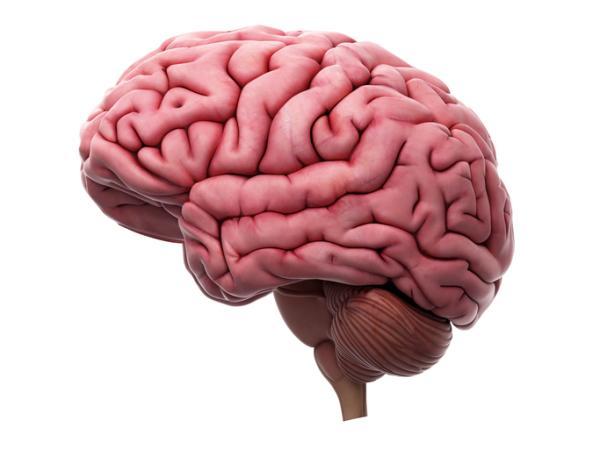
The nervous system directs all the operations of our body. This is made up of various structures and other systems that interact with each other, allowing its correct functioning.
Among these systems we find the ventricular system, which, although simple at first glance, fulfills a series of fundamental functions that directly influence the health of our brain.
Throughout this article Let’s go in depth about what the ventricular system is commenting on its development throughout the formation of the nervous system, its functions and, also, some diseases that it can present.
In the brain we find some holes, cavities called ventricles, the group of which is called the ventricular system It is a system that we could compare to pipes, a system composed of several structures in the form of cavities that connect to each other.
Although the ventricles appear empty and simple, these cavities actually fulfill fundamental functions for the nervous system, being the origin of the cerebrospinal fluid (CSF), a transparent liquid that bathes the brain and spinal cord.
Formation of the ventricular system
The ventricular system develops at the same time as the rest of the central nervous system , facilitating the circulation of CSF throughout the process. One of the first milestones in the development of this system occurs on day 26 of embryonic development (4th week), which is when the differentiation of the optic ventricle begins. Later, an outpouching begins to occur in the medial line of the midbrain, which will later constitute the cerebral or Sylvian aqueduct.
Around the 6th week, the development of the interventricular foramen begins , beginning the formation of the choroid plexuses of the lateral ventricles. From that moment on, the grooves and segmentation become more noticeable to the naked eye. After a few more weeks, the medial and lateral ventricular eminence increase in size, causing the spherical shape of the most primitive lateral ventricle to become a C. The horns of the lateral ventricles begin to become more noticeable and a small sac in the diencephalic floor, which in the future will become the third ventricle.
During the 7th and 8th weeks the end of the formation process of the ventricular system is reached It is at this moment that the horns end up becoming defined, the shape of the ventricles becoming almost definitive. The isthymic part is compressed by the cerebellum, which is still growing, and many villi extend in the midline.
Components of this system
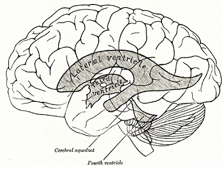
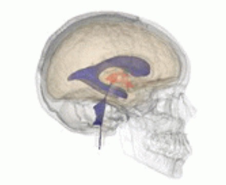
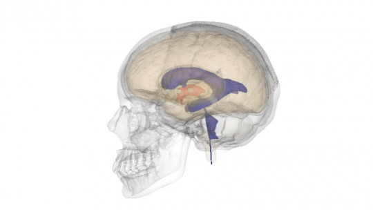
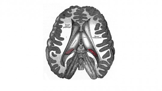
The ventricular system is made up of four ventricles, which are connected to each other through various openings and channels. Below we will see in depth what its parts are:
1. Lateral ventricles (I and II V)
The lateral ventricles are the first and second ventricles, being the largest cavities They are located deep in both cerebral hemispheres and have an anterior horn that faces the frontal lobe, and a posterior horn that faces the temporal lobe. These two ventricles are connected through the third ventricle by the interventricular foramen of Monro. Both are C-shaped and their volume increases as the years go by.
Inside each one we find the choroid plexuses. The walls and roof of both ventricles are formed by neural structures , which constitute the frontal, parietal, temporal and occipital lobes, as well as nuclei of the base and the corpus callosum. We can identify in them the frontal horn (frontal lobe), the ventricular body (frontal and parietal lobes), occipital horn (occipital lobe) and the temporal horn (temporal lobe).
2. Third ventricle (III V)
The third ventricle is a thin, flat cavity, similar in shape to that of a bird’s head It is a single cavity, smaller than the lateral ventricles and centrally located. As we have mentioned, it connects to the lateral ventricles through the foramina of Monro and to the rest of the ventricular system through the aqueduct of Silvio.
Inside we also find the choroid plexuses, specifically on its roof. The walls of this ventricle are formed by structures of the diencephalon, nuclei of the thalamus and hypothalamus. At its posterior end is the pineal gland, responsible for the production of melatonin, a hormone that regulates sleep and wake cycles.
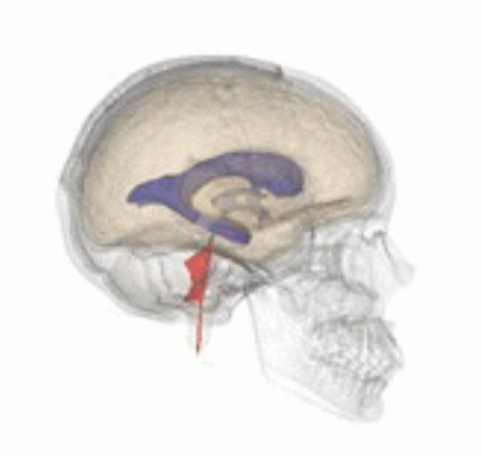
3. Fourth ventricle (IV V)
The fourth ventricle It is found occupying a space that goes from the mesencephalic aqueduct to the central canal of the upper part of the spinal cord
Its floor, that is, the surface that constitutes the base of this cavity, is formed by the rhomboid fossa and communicates with the central canal through the foramina of Luschka and Magendie, parts of which CSF exits into the subarachnoid space.. This cavity connects to the subarachnoid cisterns, which allow CSF to reach the subarachnoid space.
If we travel inside the ventricles and reach the spinal cord, we will observe that the ventricles continue through the ependymal canal This canal is a cavity that begins at the end of the fourth ventricle and runs through the spinal cord until it ends in the first vertebra of the lumbar area.
Functions of the cerebral ventricular system
Although it may seem like a very simple system due to the simple fact that it is made up of cavities, the truth is that the cerebral ventricular system performs several and very important tasks, which are the following.
1. CSF production
As we have mentioned before, The main function of the cerebral ventricles is to produce cerebrospinal fluid Likewise, it should be said that the ventricular system is not the only set of structures that form this fluid, such as the subarachnoid space, but it should be noted that the ventricles are closely involved in the production of this fluid. This substance lubricates the neural structures.
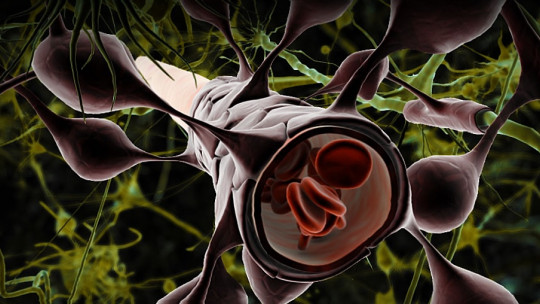
About 80% of CSF is synthesized in the choroid plexuses , and is the product resulting from the filtration of the blood that passes through them. The total volume of this fluid in an adult individual is about 150 ml. It is constantly produced and absorbed at a rate of 0.3 ml per minute, so its total volume is completely renewed about 3 times each day.
2. Brain buoyancy
CSF makes the brain float This may seem unimportant at first, but it causes the relative weight of the brain to decrease greatly, going from about 1,400 grams to about 50 grams. This means that our head “doesn’t weigh us down” as much.
3. Brain preservation
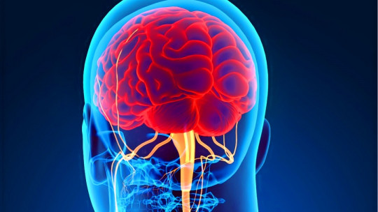
Because they produce CSF, the ventricles They help maintain internal brain homeostasis, keeping intracranial pressure constant and adequate Added to this, the ventricular system helps eliminate waste, preventing infections and fatal damage to our brain.
It is very important to understand that the brain is a very sensitive organ to any chemical and physical change within the skull, so an altered ventricular system in which not enough CSF is produced (or too much) can cause cognitive damage. , although indirectly.
As the last major function of the ventricular system, directly associated with its production of CSF, we have the fact that this fluid protects us against external agents, which could pose an infectious risk to our brain
Added to this, the CSF constitutes an effective shock absorber, making the brain trauma soften in the event that we suffer an accident, although it should be noted that it is not 100% effective and there is always the risk of injury at the cortical level. especially if the impact has been very strong.
Diseases of the ventricular system
The ventricular system can suffer from various alterations and diseases, which affect not only the health of our brain but can also cause problems for the entire organism:
1. Hydrocephalus
Hydrocephalus is caused by excessive production of CSF As this disorder progresses, intracranial pressure increases, which can lead to brain damage such as atrophy, metabolic and cognitive disorders. In the worst cases, hydrocephalus can lead to the death of the individual.
2. Ventriculitis
Ventriculitis is inflammation of the cerebral ventricles , which causes intracranial pressure to rise and also alters CSF circulation. This medical condition can be accompanied by hydrocephalus, encephalitis and inflammation of the brain.
3. Meningitis
Meningitis is inflammation of the meninges due to an infectious agent , usually fungi, viruses and bacteria. This inflammation causes an increase in intracranial pressure, making CSF circulation difficult and giving rise to different symptoms, mainly headaches, nausea, fever, sensitivity to light and in the worst cases cognitive impairment and even death.
4. Alzheimer’s disease
In Alzheimer’s disease, an cognitive impairment caused by the death of neurons , a phenomenon that increases as the same disease progresses. This causes a reduction in neuronal density, which causes the ventricles to become increasingly larger because they occupy the space left as a result of the loss of brain volume.
5. Schizophrenia
In recent years, more and more research has been carried out on the possible relationship between schizophrenia and alterations in the ventricular system It is believed that people who suffer from this psychiatric disorder could tend to have a larger dimension in the cerebral ventricles, having greater ventricular dilation and a significant cortical decrease.